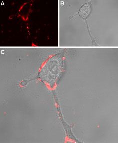
GTX54765 ICC/IF Image
ICC/IF analysis of rat PC12 cells using TRPA1 antibody at a dilution of 1:50. (B) Live view of the cells. (C) Merged image of A and B.
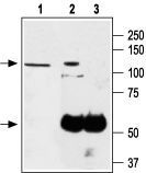
GTX54765 IP Image
IP analysis was performed with PC-12 lysates and either TRPA1 antibody (lane 2) or control IgG (lane 3). The PC-12 lysate (lane 1) and precipitates were detected by the same antibody.
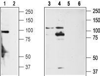
GTX54765 WB Image
WB analysis of rat DRG (lanes 1,2), non-differentiated PC12 cells (lanes 3,5) and differentiated PC12 cells (lanes 4,6) lysates using TRPA1 antibody at a dilution of 1:200 in the absence (lanes 1, 3, 4) or presence (lanes 2, 5, 6) of blocking peptide.
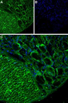
GTX54765 IHC Image
IHC analysis of rat dorsal root ganglion using TRPA1 antibody at a dilution of 1:200 (green) and DAPI (blue). (C) Merged image of A and B.