GTX49382 IHC-P Image
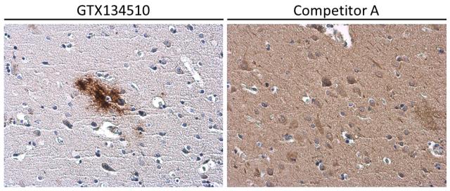
Conformation-specific beta amyloid antibody detects beta amyloid protein aggregates in the occipital lobe of human Alzheimer's disease brain by immunohistochemical analysis.Antibodies: beta amyloid (1-42) antibody ? Conformation Specific (GTX134510) diluted at 1:500, and competitor's antibody diluted at 1:500.
GTX49382 IHC-P Image
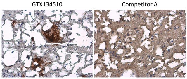
Conformation-specific beta amyloid antibody detects beta amyloid protein aggregates in the postcentral gyrus of human Alzheimer's disease brain by immunohistochemical analysis.Antibodies: beta amyloid (1-42) antibody ? Conformation Specific (GTX134510) diluted at 1:500, and competitor's antibody diluted at 1:500.
GTX49382 IHC-P Image
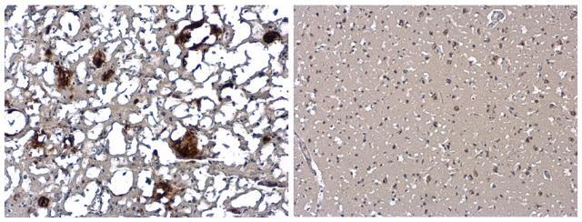
Beta amyloid (1-42) antibody (conformation specific) detects beta amyloid protein aggregates by immunohistochemical analysis in the postcentral gyrus of Alzheimer's disease brain (left), but not in normal human brain(right).Sample: Paraffin-embedded Human Brain (Alzheimer's Disease + Normal) tissue slides (GTX49382).beta amyloid protein aggregates stained by beta amyloid (1-42) antibody ? Conformation Specific (GTX49382) diluted at 1:500.
GTX49382 IHC-P Image
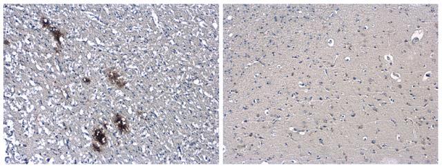
Beta amyloid (1-42) antibody (conformation specific) detects beta amyloid protein aggregates by immunohistochemical analysis in the occipital lobe of Alzheimer's disease brain (left), but not in normal human brain(right).Sample: Paraffin-embedded Human Brain (Alzheimer's Disease + Normal) tissue slides (GTX49382).beta amyloid protein aggregates stained by beta amyloid (1-42) antibody ? Conformation Specific (GTX49382) diluted at 1:500.
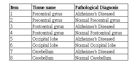
Human Matched Pair Tissue Array contains both AlzheimerÅfs disease brain tissues and normal brain tissues in different brain regions.