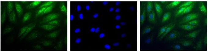
GTX30114 ICC/IF Image
Immunocytochemistry/Immunofluorescence: HIF-2 alpha Antibody (GTX30114) - Detection of HIF-2 Alpha (Green) in RCC4 cells using GTX30114. Nuclei (Blue) are counterstained with Hoechst 33258.
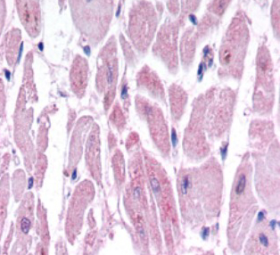
GTX30114 IHC Image
Immunohistochemistry: HIF-2 alpha Antibody (GTX30114) - Hif-2 alpha immunoreactivity in human cardiac myocytes
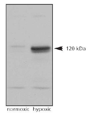
GTX30114 WB Image
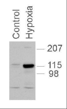
Western Blot: HIF-2 alpha Antibody (GTX30114) - Analysis on normoxic and hypoxic nuclear rat cell lysates.
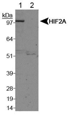
GTX30114 WB Image
Western Blot: HIF-2 alpha Antibody (GTX30114) - Analysis of HIF-2 alpha on Lane 1, Cobalt chloride treated COS7 nuclear extracts and Lane 2, Untreated COS7 nuclear extracts using GTX30114
GTX30114 WB Image
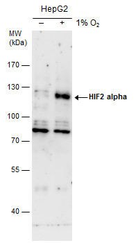
HIF2 alpha antibody detects HIF2 alpha protein by western blot analysis. Un-treated (-) and treated (+, 0.01 O2 treatment for 24h) HepG2 whole cell extracts (30 ug) were separated by 7.5% SDS-PAGE, and the membrane was blotted with HIF2 alpha antibody (GTX30114) diluted by 1:500. The HRP-conjugated anti-rabbit IgG antibody (GTX213110-01) was used to detect the primary antibody.
GTX30114 WB Image
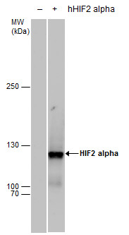
HIF2 alpha antibody detects HIF2 alpha protein by western blot analysis. Non-transfected (-) and HIF2A-transfected (+, including Myc-DDK-tag) 293T whole cell extracts (30 ug) were separated by 5% SDS-PAGE, and the membrane was blotted with HIF2 alpha antibody (GTX30114) diluted by 1:1000. The HRP-conjugated anti-rabbit IgG antibody (GTX213110-01) was used to detect the primary antibody.
GTX30114 WB Image
GTX30114 utilized in western blot with PC12 nuclear extracts, 1:1,000, 5 second exposure.
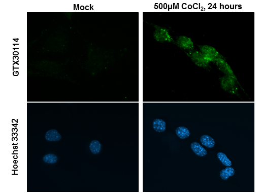
GTX30114 ICC/IF Image
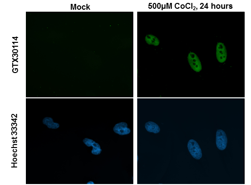
HIF2 alpha antibody detects HIF2 alpha protein at nucleus by immunofluorescent analysis.Sample: HeLa cells were fixed in 4% paraformaldehyde at RT for 15 min.Green: HIF2 alpha stained by HIF2 alpha antibody (GTX30114) diluted at 1:200.Blue: Hoechst 33342 staining.
GTX30114 ICC/IF Image
HIF2 alpha antibody detects HIF2 alpha protein at nucleus by immunofluorescent analysis.Sample: NIH-3T3 cells were fixed in 4% paraformaldehyde at RT for 15 min.Green: HIF2 alpha stained by HIF2 alpha antibody (GTX30114) diluted at 1:200.Blue: Hoechst 33342 staining.