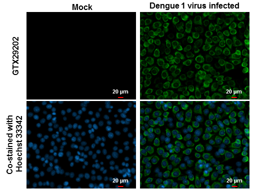
GTX29202 ICC/IF Image
Dengue virus 1, 2, 3 & 4 antibody [D1-11(3)] detects Dengue virus 1 protein at cytoplasm by immunofluorescent analysis.
Samples: BHK-21 cells mock (left) and infected with Dengue virus 1 (right) were fixed in MeOH.
Green: Dengue virus 1 protein stained by Dengue virus 1, 2, 3 & 4 antibody [D1-11(3)] (GTX29202) diluted at 1:500.
Blue: Hoechst 33342 staining.
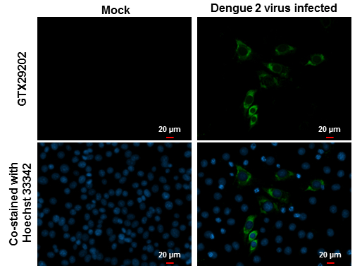
GTX29202 ICC/IF Image
Dengue virus 1, 2, 3 & 4 antibody [D1-11(3)] detects Dengue virus 2 protein at cytoplasm by immunofluorescent analysis.
Samples: BHK-21 cells mock (left) and infected with Dengue virus 2 (right) were fixed in MeOH.
Green: Dengue virus 2 protein stained by Dengue virus 1, 2, 3 & 4 antibody [D1-11(3)] (GTX29202) diluted at 1:500.
Blue: Hoechst 33342 staining.
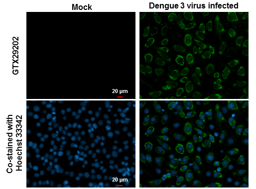
GTX29202 ICC/IF Image
Dengue virus 1, 2, 3 & 4 antibody [D1-11(3)] detects Dengue virus 3 protein at cytoplasm by immunofluorescent analysis.
Samples: BHK-21 cells mock (left) and infected with Dengue virus 3 (right) were fixed in MeOH.
Green: Dengue virus 3 protein stained by Dengue virus 1, 2, 3 & 4 antibody [D1-11(3)] (GTX29202) diluted at 1:500.
Blue: Hoechst 33342 staining.
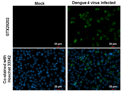
GTX29202 ICC/IF Image
Dengue virus 1, 2, 3 & 4 antibody [D1-11(3)] detects Dengue virus 4 protein at cytoplasm by immunofluorescent analysis.
Samples: BHK-21 cells mock (left) and infected with Dengue virus 4 (right) were fixed in MeOH.
Green: Dengue virus 4 protein stained by Dengue virus 1, 2, 3 & 4 antibody [D1-11(3)] (GTX29202) diluted at 1:500.
Blue: Hoechst 33342 staining.