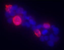
Immunohistochemistry using anti-Gli-1 antibody shows detection of mouse Gli-1 present in transfected 293T cells (red). HEK293T cells were transiently transfected with Gli-1 (murine). Anti-Gli-1 antiserum (rabbit) was added 1:400, followed by a fluorescent labeled anti-rabbit IgG secondary. Personal communication, Tom Curran, Children’s Hospital of Philadelphia, Philadelphia, PA.
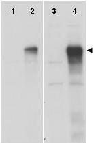
Western blot using GeneTex anti-Gli-1 antibody (GTX27523) shows detection of a band at ~150 kDa (arrowhead) corresponding to human Gli-1 present in transfected 293T cell lysates (lanes 2 and 4). Mock 293T cell lysates with vector only show no staining (lanes 1 and 3). Lysates were separated by SDS-PAGE and transferred to nitrocellulose. After blocking the membrane was probed with the primary antibody diluted to 1:8,000 (lanes 1 and 2) or 1:4,000 (lanes 3 and 4). Molecular weight estimation was made by comparison to MW markers. Personal communication, Hiro Kimura, St. Jude Children's Research Hospital, Memphis, TN.
Western blot using GeneTex anti-Gli-1 antibody (GTX27523) shows detection of a band at ~150 kDa (arrowhead) corresponding to human Gli-1 present in transfected 293T cell lysates (lanes 2 and 4). Mock 293T cell lysates with vector only show no staining (lanes 1 and 3).? Lysates were separated by SDS-PAGE and transferred to nitrocellulose.?After blocking the membrane was probed with the primary antibody diluted to 1:8,000 (lanes 1 and 2) or 1:4,000 (lanes 3 and 4). Molecular weight estimation was made by comparison to MW markers.? Personal communication, Hiro Kimura, St. Jude Children's Research Hospital, Memphis, TN.