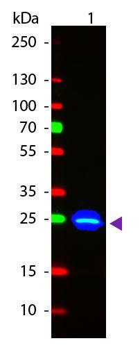
GTX26662 WB Image
Western Blot of Goat anti-GFP Fluorescein Conjugated Antibody (GTX26662). Lane 1: GFP. Load: 50 ng per lane. Primary antibody: None. Secondary antibody: Fluorescein goat secondary antibody at 1:1,000 for 60 min at RT. Block for 30 min at RT. Predicted/Observed size: 28 kDa, 33 kDa for GFP. Other band(s): None.
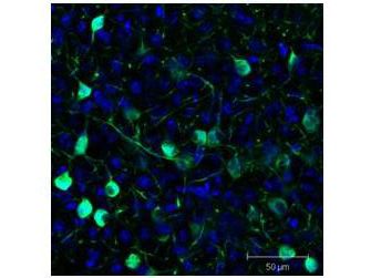
GTX26662 IHC-Fr Image
Immunofluorescence Microscopy of GFP-GOAT-Antibody. Tissue: Sf-1:Cre mice crossed to the Z/EG reporter line. Mouse brain (coronal view, 20X magnification). Fixation: 4%PFA/PBS with o/n fixation, and subsequently transferred to a 30% sucrose solution. Antigen retrieval: frozen in OCT freezing medium (Sakura) and cryostat sectioned at 40 microns. Primary antibody: Goat anti-GFP (GTX26662) was used at 1:500 dilution in free floating imunnohistochemistry to detect GFP. Secondary antibody: Fluorchrome conjugated Anti-goat IgG secondary antibody was used for detection at 1:500 at 1:10,000 for 45 min at RT. Localization: Sf-1+ neurons and their processes of the ventromedial nucleus of the hypothalamus. Staining: eGFP as green fluorescent signal and sections were counterstained with DAPI.