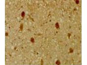
GTX26545 IHC-P Image
Immunohistochemical staining of mouse tissue using anti-pRb2/p130 antiserum.? The staining shows the location of pRb2/p130 in developing mouse tissue.? Other detection systems should yield similar results. Sections were cut at 5-7 um, mounted on glass and dried overnight at 37üŗC. ?All sections were deparaffinized in xylene, rehydrated through a graded alcohol series and washed in phosphate-buffered saline (PBS). ?PBS was used for all subsequent washes and for antiserum dilution. ?Tissue sections were quenched sequentially in 0.5% hydrogen peroxide and blocked with diluted 10% normal goat anti-rabbit serum. ?Slides were incubated at 20üŗ C for 1 h with rabbit anti-pRb2/p130 (1:500) dilution, washed, and then reacted with diluted goat anti-rabbit biotinylated antibody for 30 min. ?Slides were then reacted with streptavidin-peroxidase conjugate for 30 min at 20üŗ C.? Diaminobenzidine was used as the final chromogen. ?Negative controls for each tissue section were prepared by substituting the primary antiserum with pre-immune serum.
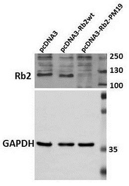
GTX26545 WB Image
Western Blot of Rabbit Anti-Rb2 p130 Antibody. Lane 1: HEK 293 pcDNA3. Lane 2: HEK 293 pcDNA3-Rb2wt. Lane 3: HEK 293 pcDNA3-Rb2-PM19. Load: 30 ug per lane. Primary antibody: Anti-Rb2 antibody at 1:250 for overnight at 4üŗC. Secondary antibody: IRDye800? rabbit secondary antibody at 1:10,000 for 45 min at RT. Block: 5% BLOTTO overnight at 4üŗC. Predicted/Observed size: 130 kDa for p130/Rb2.