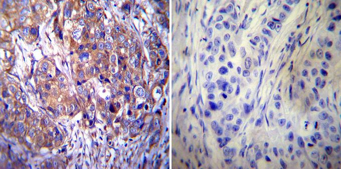
GTX22731 IHC-P Image
Immunohistochemistry was performed on cancer biopsies of deparaffinized human breast carcinoma tissue. To expose target proteins, heat induced antigen retrieval was performed using 10mM sodium citrate (pH6.0) buffer, microwaved for 8-15 minutes. Following antigen retrieval tissues were blocked in 3% BSA-PBS for 30 minutes at room temperature. Tissues were then probed at a dilution of 1:100 with or without Clathrin heavy chain antibody [X22] overnight at 4üŗC in a humidified chamber. Tissues were washed extensively with PBST and endogenous peroxidase activity was quenched with a peroxidase suppressor. Detection was performed using a biotin-conjugated secondary antibody and SA-HRP, followed by colorimetric detection using DAB. Tissues were counterstained with hematoxylin and prepped for mounting.

GTX22731 ICC/IF Image
Immunofluorescent analysis of Clathrin heavy chain in HeLa cells. Clathrin heavy chain staining (green), F-Actin staining with Phalloidin (red) and nuclei with DAPI (blue) is shown. Cells were grown on slides and fixed with formaldehyde prior to staining. Cells were probed without (control) or with Clathrin heavy chain antibody [X22] at a dilution of 1:200 over night at 4 ?C, washed with PBS and incubated with a proper secondary antibody. Images were taken at 60X magnification.