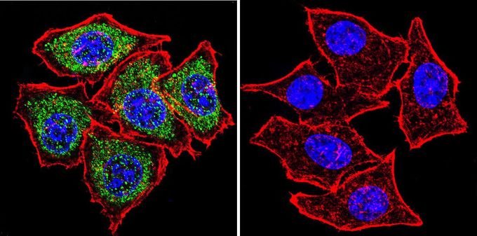
GTX15779 ICC/IF Image
Immunofluorescent analysis of PLK1 in HeLa cells. PLK1 staining (green), F-Actin staining with Phalloidin (red) and nuclei with DAPI (blue) is shown. Cells were grown on slides and fixed with formaldehyde prior to staining. Cells were probed without (control) or with PLK1 antibody [13E8] at a dilution of 1:20 over night at 4üŗC, washed with PBS and incubated with a proper secondary antibody. Images were taken at 60X magnification.
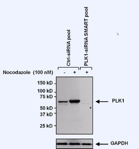
GTX15779 WB Image
Western blot analysis of PLK1 in 25ug of untreated or Nocodazole (100nM, 48 hours) treated U2OS lysate from non-targeting control or PLK1 siRNA transfected U2OS cells. Proteins were transferred to a PVDF membrane and blocked with 5% Milk/TBST for at least 1 hour. Membranes were probed with PLK1 antibody [13E8] at a dilution of 1:1000 overnight at 4üŗC on a rocking platform. Membranes were washed in TBS-0.1%Tween 20 and probed with a HRP-conjugated secondary antibody. Membranes were washed and chemiluminescent detection was performed.

GTX15779 IHC-P Image
Immunohistochemistry was performed on normal deparaffinized human colon tissue. To expose target proteins, heat induced antigen retrieval was performed using 10mM sodium citrate (pH6.0) buffer, microwaved for 8-15 minutes. Following antigen retrieval tissues were blocked in 3% BSA-PBS for 30 minutes at room temperature. Tissues were then probed at a dilution of 1:200 with or without PLK1 antibody [13E8] overnight at 4üŗC in a humidified chamber. Tissues were washed extensively with PBST and endogenous peroxidase activity was quenched with a peroxidase suppressor. Detection was performed using a biotin-conjugated secondary antibody and SA-HRP, followed by colorimetric detection using DAB. Tissues were counterstained with hematoxylin and prepped for mounting.