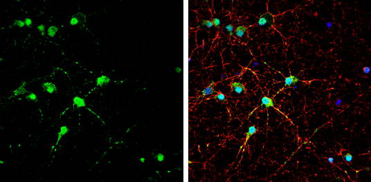
GTX134100 ICC/IF Image
EAAT4 antibody detects EAAT4 protein by immunofluorescent analysis.Sample: DIV10 rat E18 primary cortical neuron cells were fixed in 4% paraformaldehyde at RT for 15 min.Green: EAAT4 stained by EAAT4 antibody (GTX134100) diluted at 1:500.Red: alpha Tubulin, stained by alpha Tubulin antibody [GT114] (GTX628802) diluted at 1:500.Blue: Fluoroshield with DAPI (GTX30920).
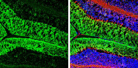
GTX134100 IHC-Fr Image
EAAT4 antibody detects EAAT4 protein by immunohistochemical analysis.Sample: Frozen-sectioned mouse cerebellum.Green: EAAT4 stained by EAAT4 antibody (GTX134100) diluted at 1:250.Red: NF-H, stained by NF-H antibody [GT114] (GTX634289) diluted at 1:500.Blue: Fluoroshield with DAPI (GTX30920).
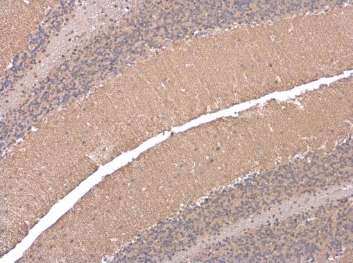
GTX134100 IHC-P Image
EAAT4 antibody detects EAAT4 protein at cell membrane by immunohistochemical analysis.Sample: Paraffin-embedded rat brain.EAAT4 stained by EAAT4 antibody (GTX134100) diluted at 1:500.