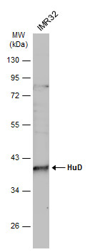
GTX134099 WB Image
Whole cell extract (30 ug) was separated by 10% SDS-PAGE, and the membrane was blotted with HuD antibody (GTX134099) diluted at 1:500. The HRP-conjugated anti-rabbit IgG antibody (GTX213110-01) was used to detect the primary antibody.
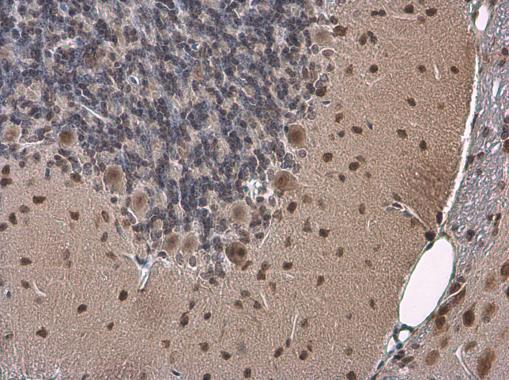
GTX134099 IHC-P Image
HuD antibody detects HuD protein at cytoplasm and nucleus by immunohistochemical analysis.Sample: Paraffin-embedded mouse brain.HuD stained by HuD antibody (GTX134099) diluted at 1:500.
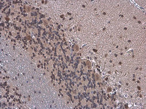
GTX134099 IHC-P Image
HuD antibody detects HuD protein at cytoplasm and nucleus by immunohistochemical analysis.Sample: Paraffin-embedded rat brain.HuD stained by HuD antibody (GTX134099) diluted at 1:500.
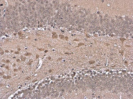
GTX134099 IHC-P Image
HuD antibody detects HuD protein at cytoplasm and nucleus by immunohistochemical analysis.Sample: Paraffin-embedded mouse hippocampus.HuD stained by HuD antibody (GTX134099) diluted at 1:500.