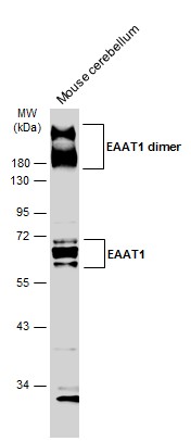
GTX134059 WB Image
Mouse tissue extract (50 ug) was separated by 10% SDS-PAGE, and the membrane was blotted with EAAT1 antibody (GTX134059) diluted at 1:1000. The HRP-conjugated anti-rabbit IgG antibody (GTX213110-01) was used to detect the primary antibody, and the signal was developed with Trident ECL plus-Enhanced.
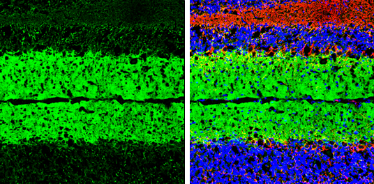
GTX134059 IHC-Fr Image
EAAT1 antibody detects EAAT1 protein by immunohistochemical analysis.Sample: Frozen-sectioned mouse cerebellum.Green: EAAT1 stained by EAAT1 antibody (GTX134059) diluted at 1:250.Red: NF-H, stained by NF-H antibody [GT114] (GTX634289) diluted at 1:500.Blue: Fluoroshield with DAPI (GTX30920).
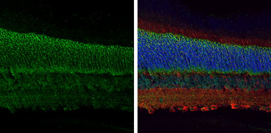
GTX134059 IHC-P Image
EAAT1 antibody detects EAAT1 protein at cell membrane by immunohistochemical analysis.Sample: Paraffin-embedded mouse eye.Green: EAAT1 stained by EAAT1 antibody (GTX134059) diluted at 1:250.Red: beta Tubulin 3/ Tuj1, a cytoplasm marker, stained by beta Tubulin 3/ Tuj1 antibody [GT1338] (GTX631831) diluted at 1:500.Blue: Fluoroshield with DAPI (GTX30920).
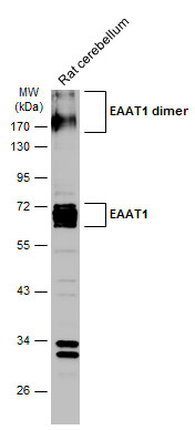
GTX134059 WB Image
Rat tissue extract (50 ug) was separated by 10% SDS-PAGE, and the membrane was blotted with EAAT1 antibody (GTX134059) diluted at 1:1000. The HRP-conjugated anti-rabbit IgG antibody (GTX213110-01) was used to detect the primary antibody.