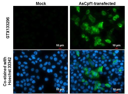
GTX133296 ICC/IF Image
CPF1 antibody detects CPF1 protein at cytoplasm by immunofluorescent analysis.
Sample: 293T cells were fixed in 4% paraformaldehyde at RT for 15 min.
Green: CPF1 protein stained by CPF1 antibody (GTX133296) diluted at 1:500.
Blue: Hoechst 33342 staining.
Scale bar = 10 um.
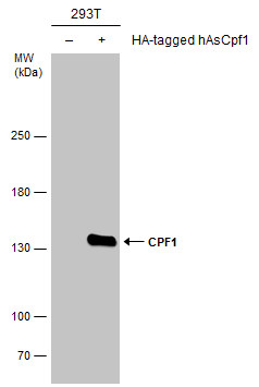
GTX133296 WB Image
Non-transfected (?) and transfected (+) 293T whole cell extracts (5 ug) were separated by 5% SDS-PAGE, and the membrane was blotted with CPF1 antibody (GTX133296) diluted at 1:5000.