GTX133148 IHC-Fr Image
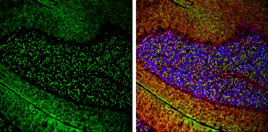
VGluT1 antibody detects VGluT1 protein expression by immunohistochemical analysis.
Sample: Frozen-sectioned adult mouse cerebellum.
Green: VGluT1 protein stained by VGluT1 antibody (GTX133148) diluted at 1:250.
Red: beta Tubulin 3/ TUJ1, stained by beta Tubulin 3/ TUJ1 antibody [GT11710] (GTX631836) diluted at 1:500.
Blue: Fluoroshield with DAPI (GTX30920).
GTX133148 ICC/IF Image
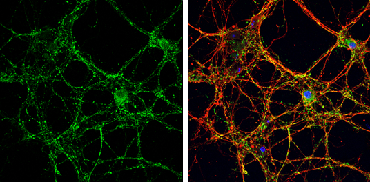
VGluT1 antibody detects VGluT1 protein by immunofluorescent analysis.
Sample: DIV14 rat E18 primary cortical neurons were fixed in 4% paraformaldehyde at RT for 15 min.
Green: VGluT1 protein stained by VGluT1 antibody (GTX133148) diluted at 1:500.
Red: beta Tubulin 3/ Tuj1, stained by beta Tubulin 3/ Tuj1 antibody [GT1338] (GTX631831) diluted at 1:500.
Blue: Fluoroshield with DAPI (GTX30920).
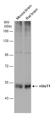
GTX133148 WB Image
Various tissue extracts (50 ug) were separated by 7.5% SDS-PAGE, and the membrane was blotted with vGluT1 antibody (GTX133148) diluted at 1:1000.
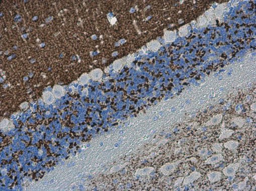
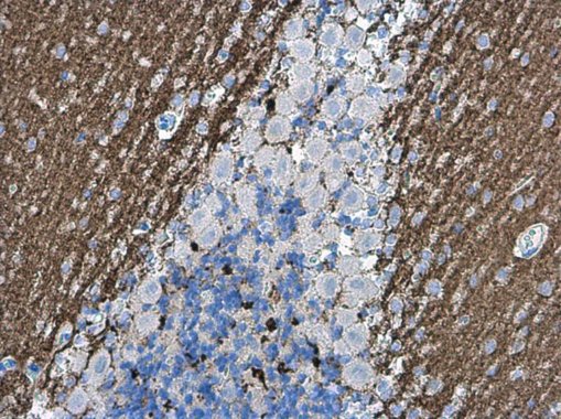
GTX133148 IHC-P Image
VGluT1 antibody detects VGluT1 protein at cell membrane in mouse brain by immunohistochemical analysis.
Sample: Paraffin-embedded mouse brain.
VGluT1 antibody (GTX133148) diluted at 1:500.
GTX133148 IHC-P Image
VGluT1 antibody detects VGluT1 protein at cell membrane in rat brain by immunohistochemical analysis.
Sample: Paraffin-embedded rat brain.
VGluT1 antibody (GTX133148) diluted at 1:500.