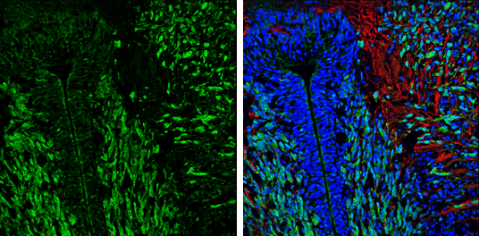
GTX133108 IHC-Fr Image
Myelin Basic Protein antibody detects Myelin Basic Protein protein expression by immunohistochemical analysis.
Sample: Frozen sectioned E13.5 Rat brain.
Green: Myelin Basic Protein protein stained by Myelin Basic Protein antibody (GTX133108) diluted at 1:250.
Red: beta Tubulin 3/ TUJ1, a mature neuron marker, stained by beta Tubulin 3/ TUJ1 antibody [GT11710] (GTX631836) diluted at 1:500.
Blue: Fluoroshield with DAPI (GTX30920).
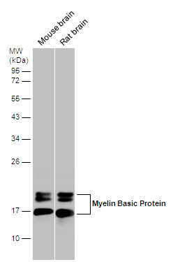
GTX133108 WB Image
Various tissue extracts (50 ug) were separated by 12% SDS-PAGE, and the membrane was blotted with Myelin Basic Protein antibody (GTX133108) diluted at 1:10000.
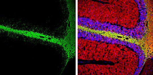
GTX133108 IHC-Fr Image
Myelin Basic Protein antibody detects Myelin Basic Protein expression by immunohistochemical analysis.
Sample: Frozen-sectioned adult mouse cerebellum.
Green: Myelin Basic Protein stained by Myelin Basic Protein antibody (GTX133108) diluted at 1:250.
Red: beta Tubulin 3/ TUJ1, stained by beta Tubulin 3/ TUJ1 antibody [GT11710] (GTX631836) diluted at 1:500.
Blue: Fluoroshield with DAPI (GTX30920).
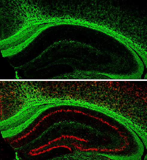
GTX133108 IHC-Fr Image
Myelin Basic Protein antibody detects Myelin Basic protein expression by immunohistochemical analysis.
Sample: Frozen-sectioned adult mouse hippocampus.
Green: Myelin Basic protein stained by Myelin Basic Protein antibody (GTX133108) diluted at 1:250.
Red: NeuN, stained by NeuN antibody [2Q158] (GTX30773) diluted at 1:500.
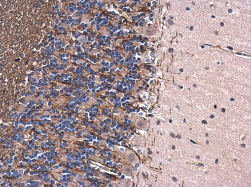
GTX133108 IHC-P Image
Myelin Basic Protein antibody detects Myelin Basic Protein protein at cytoplasm in rat brain by immunohistochemical analysis.
Sample: Paraffin-embedded rat brain.
Myelin Basic Protein antibody (GTX133108) diluted at 1:500.
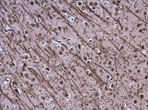
GTX133108 IHC-P Image
Myelin Basic Protein antibody detects Myelin Basic Protein protein at cytoplasm in rat brain by immunohistochemical analysis.
Sample: Paraffin-embedded rat brain.
Myelin Basic Protein antibody (GTX133108) diluted at 1:500.