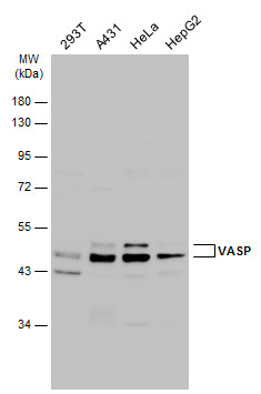
GTX132983 WB Image
Various whole cell extracts (30 ug) were separated by 10% SDS-PAGE, and the membrane was blotted with VASP antibody (GTX132983) diluted at 1:1000. The HRP-conjugated anti-rabbit IgG antibody (GTX213110-01) was used to detect the primary antibody.
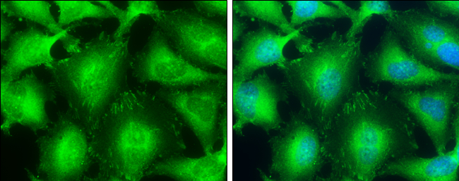
GTX132983 ICC/IF Image
VASP antibody detects VASP protein at focal adhesion by immunofluorescent analysis.
Sample: HeLa cells were fixed in 4% paraformaldehyde at RT for 15 min.
Green: VASP protein stained by VASP antibody (GTX132983) diluted at 1:500.
Blue: Hoechst 33342 staining.
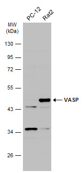
GTX132983 WB Image
Various whole cell extracts (30 ug) were separated by 10% SDS-PAGE, and the membrane was blotted with VASP antibody (GTX132983) diluted at 1:1000. The HRP-conjugated anti-rabbit IgG antibody (GTX213110-01) was used to detect the primary antibody.
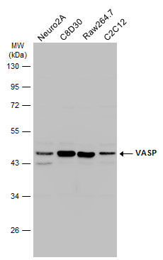
GTX132983 WB Image
Various whole cell extracts (30 ug) were separated by 10% SDS-PAGE, and the membrane was blotted with VASP antibody (GTX132983) diluted at 1:1000. The HRP-conjugated anti-rabbit IgG antibody (GTX213110-01) was used to detect the primary antibody.