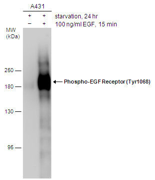
GTX132810 WB Image
Untreated (?) and treated (+) A431 whole cell extracts (20 ug) were separated by 7.5% SDS-PAGE, and the membrane was blotted with Phospho-EGF Receptor (Tyr1068) antibody (GTX132810) diluted at 1:2000.
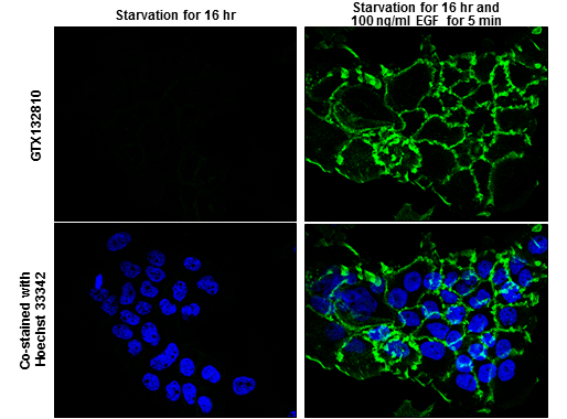
GTX132810 ICC/IF Image
EGFR (phospho Tyr1068) antibody detects EGFR (phospho Tyr1068) protein at cell membrane by immunofluorescent analysis.
Sample: A431 cells were fixed in 4% paraformaldehyde at RT for 15 min.
Green: EGFR (phospho Tyr1068) protein stained by EGFR (phospho Tyr1068) antibody (GTX132810) diluted at 1:500.
Blue: Hoechst 33342 staining.
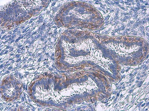
GTX132810 IHC-P Image
EGFR (phospho Tyr1068) antibody detects EGFR (phospho Tyr1068) protein at cell membrane in human breast carcinoma by immunohistochemical analysis.
Sample: Paraffin-embedded human breast carcinoma.
EGFR (phospho Tyr1068) antibody (GTX132810) diluted at 1:500.