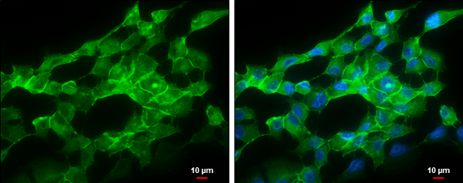
GTX132646 ICC/IF Image
pan Cadherin antibody detects pan Cadherin protein at cell membrane by immunofluorescent analysis.
Sample: SK-N-AS cells were fixed in ice-cold MeOH for 5 min.
Green: pan Cadherin protein stained by pan Cadherin antibody (GTX132646) diluted at 1:500.
Blue: Hoechst 33342 staining.
Scale bar = 10 um.
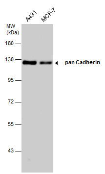
GTX132646 WB Image
Various whole cell extracts (30 ug) were separated by 7.5% SDS-PAGE, and the membrane was blotted with pan Cadherin antibody (GTX132646) diluted at 1:5000.
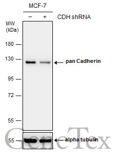
GTX132646 WB Image
Non-transfected (?) and transfected (+) MCF-7 whole cell extracts (30 ug) were separated by 7.5% SDS-PAGE, and the membrane was blotted with pan Cadherin antibody (GTX132646) diluted at 1:4000.