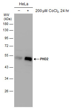
GTX132293 WB Image
Untreated (?) and treated (+) HeLa whole cell extracts (30 ug) were separated by 10% SDS-PAGE, and the membrane was blotted with PHD2 antibody (GTX132293) diluted at 1:2000.
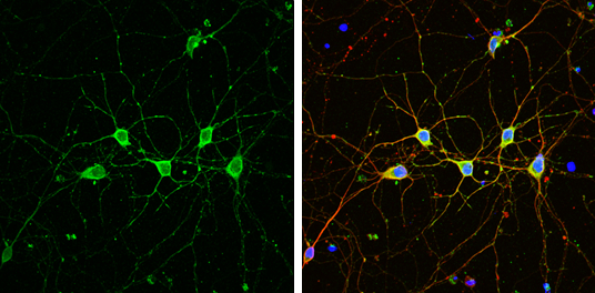
GTX132293 ICC/IF Image
PHD2 antibody detects PHD2 protein by immunofluorescent analysis.Sample: DIV9 rat E18 primary cortical neuron cells were fixed in 4% paraformaldehyde at RT for 15 min.Green: PHD2 stained by PHD2 antibody (GTX132293) diluted at 1:500.Red: beta Tubulin 3/ Tuj1, stained by beta Tubulin 3/ Tuj1 antibody [GT1338] (GTX631831) diluted at 1:500.Blue: Fluoroshield with DAPI (GTX30920).
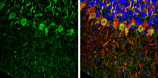
GTX132293 IHC-Fr Image
PHD2 antibody detects PHD2 Protein expression by immunohistochemical analysis.
Sample: Frozen-sectioned adult mouse cerebellum.
Green: PHD2 stained by PHD2 antibody (GTX132293) diluted at 1:250.
Red: NF-H, stained by NF-H antibody [GT114] (GTX634289) diluted at 1:500.
Blue: Fluoroshield with DAPI (GTX30920).