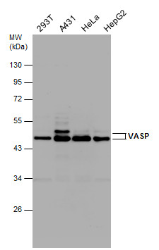
GTX132176 WB Image
Various whole cell extracts (30 ug) were separated by 10% SDS-PAGE, and the membrane was blotted with VASP antibody (GTX132176) diluted at1:1000.
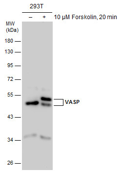
GTX132176 WB Image
Untreated (?) and treated (+) 293T whole cell extracts (30 ug) were separated by 10% SDS-PAGE, and the membrane was blotted with VASP antibody (GTX132176) diluted at 1:2000.
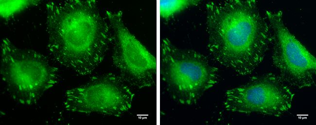
GTX132176 ICC/IF Image
VASP antibody detects VASP protein at cytoplasm by immunofluorescent analysis.
Sample: HeLa cells were fixed in 4% paraformaldehyde at RT for 10 min.
Green: VASP protein stained by VASP antibody (GTX132176) diluted at 1:500.
Blue: Hoechst 33342 staining.
Scale bar = 10 um.
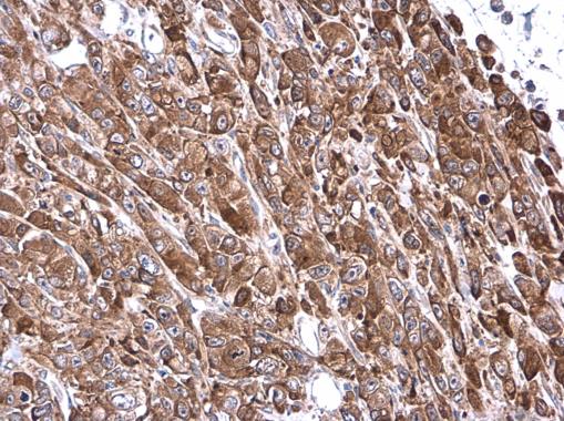
GTX132176 IHC-P Image
VASP antibody detects VASP protein at cytoplasm in MDA-MB-231 xenograft by immunohistochemical analysis.
Sample: Paraffin-embedded MDA-MB-231 xenograft.
VASP antibody (GTX132176) diluted at 1:500.