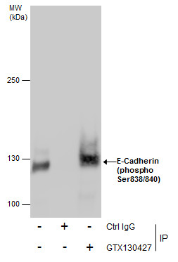
GTX130427 IP Image
Immunoprecipitation of E-Cadherin (phospho Ser838/840) protein from MCF-7 whole cell extracts using 5 ug of E-Cadherin (phospho Ser838/840) antibody (GTX130427).
Western blot analysis was performed using E-Cadherin (phospho Ser838/840) antibody (GTX130427).
EasyBlot anti-Rabbit IgG (GTX221666-01) was used as a secondary reagent.
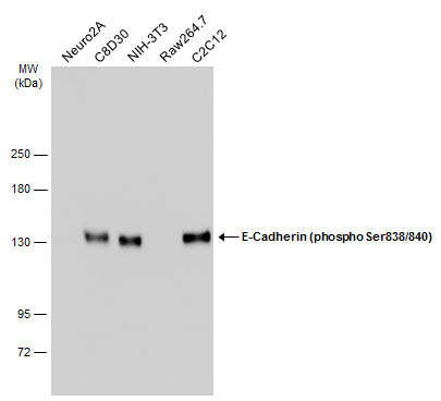
GTX130427 WB Image
Various whole cell extracts (30 ug) were separated by 5% SDS-PAGE, and the membrane was blotted with E-Cadherin (phospho Ser838/840) antibody (GTX130427) diluted at 1:1000. The HRP-conjugated anti-rabbit IgG antibody (GTX213110-01) was used to detect the primary antibody.
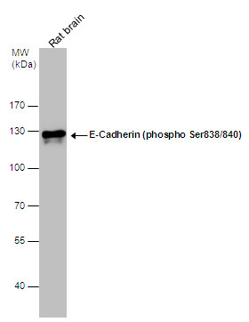
GTX130427 WB Image
E-Cadherin (phospho Ser838/840) antibody detects E-Cadherin (phospho Ser838/840) protein by western blot analysis. Rat tissue extracts (50 ug) was separated by 7.5% SDS-PAGE, and the membrane was blotted with E-Cadherin (phospho Ser838/840) antibody (GTX130427) diluted at 1:10000.
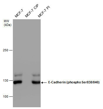
GTX130427 WB Image
E-Cadherin (phospho Ser838/840) antibody detects E-Cadherin (phospho Ser838/840) protein by western blot analysis. MCF-7 whole cell extract (30 ug) with the indicated treatments were separated by 5% SDS-PAGE, and the membrane was blotted with E-Cadherin (phospho Ser838/840) antibody (GTX130427) at a dilution of 1:10000.