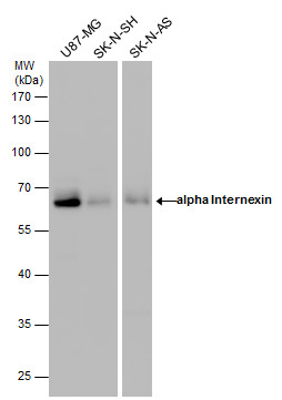
GTX130053 WB Image
alpha Internexin antibody detects alpha Internexin protein by western blot analysis. Various whole cell extracts (30 ug) were separated by 10% SDS-PAGE, and the membrane was blotted with alpha Internexin antibody (GTX130053) diluted by 1:1000.
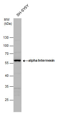
GTX130053 WB Image
alpha Internexin antibody detects alpha Internexin protein by western blot analysis. Whole cell extracts (30 ug) was separated by 10% SDS-PAGE, and the membrane was blotted with alpha Internexin antibody (GTX130053) diluted by 1:1000.
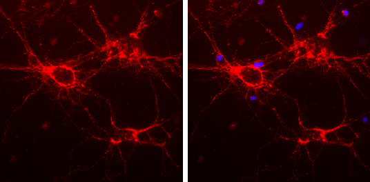
GTX130053 ICC/IF Image
alpha Internexin antibody detects alpha Internexin protein expression by immunofluorescent analysis.
Sample: Cultured rat E18 primary cortical neuron, DIV 8. Cells were fixed in 4% paraformaldehyde at RT for 15 min.
Red: alpha Internexin protein stained by alpha Internexin antibody (GTX130053) diluted at 1:250.
Blue: Fluoroshield with DAPI (GTX30920).
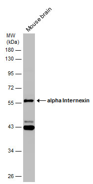
GTX130053 WB Image
Mouse tissue extract (50 ug) was separated by 10% SDS-PAGE, and the membrane was blotted with alpha Internexin antibody (GTX130053) diluted at 1:1000.
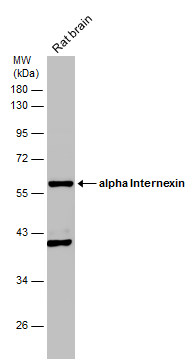
GTX130053 WB Image
Rat tissue extract (50 ug) was separated by 10% SDS-PAGE, and the membrane was blotted with alpha Internexin antibody (GTX130053) diluted at 1:1000.
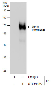
GTX130053 IP Image
Immunoprecipitation of alpha Internexin protein from U87-MG whole cell extracts using 5 ug of alpha Internexin antibody (GTX130053).
Western blot analysis was performed using alpha Internexin antibody (GTX130053).
EasyBlot anti-Rabbit IgG (GTX221666-01) was used as a secondary reagent.