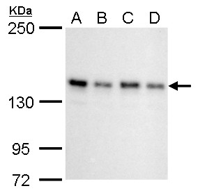
GTX129156 WB Image
SIN3A antibody detects SIN3A protein by western blot analysis.
A 293T whole cell lysate/extract
B A431 whole cell lysate/extract
C HeLa whole cell lysate/extract
D HepG2 whole cell lysate/extract
5% SDS-PAGE
SIN3A antibody (GTX129156) dilution: 1:1000
The HRP-conjugated anti-rabbit IgG antibody (GTX213110-01) was used to detect the primary antibody.
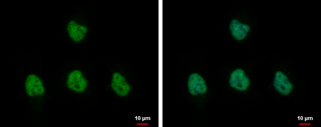
GTX129156 ICC/IF Image
SIN3A antibody detects SIN3A protein at nucleus by immunofluorescent analysis.
Sample: HeLa cells were fixed in 4% paraformaldehyde at RT for 15 min.
Green: SIN3A protein stained by SIN3A antibody (GTX129156) diluted at 1:1000.
Blue: Hoechst 33342 staining.
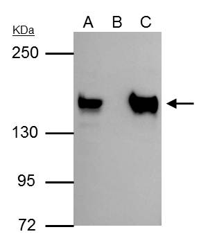
GTX129156 IP Image
SIN3A antibody immunoprecipitates SIN3A protein in IP experiments. IP Sample: 293T whole cell lysate/extract A : 30 ug whole cell lysate/extract of SIN3A protein expressing 293T cells B : Control with 3 ug of pre-immune rabbit IgG C : Immunoprecipitation of SIN3A by 3 ug of SIN3A antibody (GTX129156) 5% SDS-PAGE The immunoprecipitated SIN3A protein was detected by SIN3A antibody (GTX129156) diluted at 1 : 1000. EasyBlot anti-rabbit IgG (HRP) (GTX221666-01) was used as a secondary reagent.
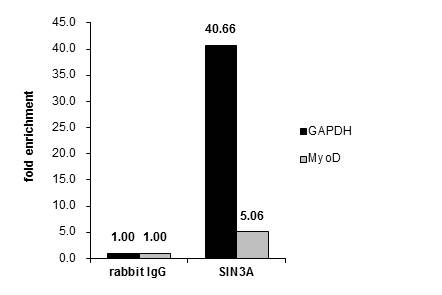
GTX129156 ChIP assay Image
Cross-linked ChIP was performed with MCF-7 chromatin extract treated with B-estradiol (10 nM for 45 min) and 5 ug of either control rabbit IgG or anti-SIN3A antibody. The precipitated DNA was detected by PCR with primer set targeting to GAPDH or MyoD.
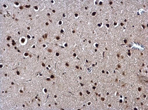
GTX129156 IHC-P Image
SIN3A antibody detects SIN3A protein at nucleus on rat fore brain by immunohistochemical analysis.
Sample: Paraffin-embedded rat fore brain.
SIN3A antibody (GTX129156) dilution: 1:500.
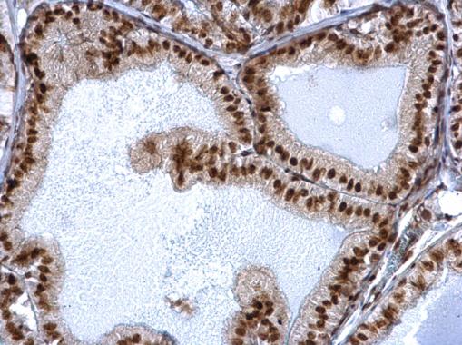
GTX129156 IHC-P Image
SIN3A antibody detects SIN3A protein at nucleus on mouse prostate by immunohistochemical analysis.
Sample: Paraffin-embedded mouse prostate.
SIN3A antibody (GTX129156) dilution: 1:500.
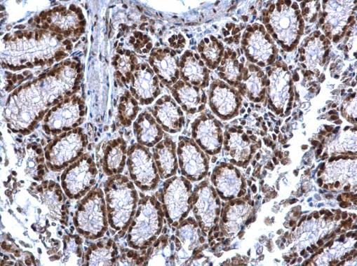
GTX129156 IHC-P Image
SIN3A antibody detects SIN3A protein at nucleus on mouse colon by immunohistochemical analysis.
Sample: Paraffin-embedded mouse colon.
SIN3A antibody (GTX129156) dilution: 1:500.
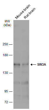
GTX129156 WB Image
Various tissue extracts (50 ug) were separated by 5% SDS-PAGE, and the membrane was blotted with SIN3A antibody (GTX129156) diluted at 1:1000. The HRP-conjugated anti-rabbit IgG antibody (GTX213110-01) was used to detect the primary antibody.