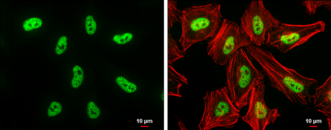
GTX129127 ICC/IF Image
DNMT3B antibody detects DNMT3B protein at nucleus by immunofluorescent analysis.
Sample: HeLa cells were fixed in 4% paraformaldehyde at RT for 15 min.
Green: DNMT3B protein stained by DNMT3B antibody (GTX129127) diluted at 1:200.
Red: phalloidin, a cytoskeleton marker, diluted at 1:50.
Scale bar = 10 um.
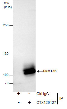
GTX129127 IP Image
Immunoprecipitation of DNMT3B protein from K562 nuclear extracts using 5 ug of DNMT3B antibody (GTX129127).
Western blot analysis was performed using DNMT3B antibody (GTX129127).
EasyBlot anti-Rabbit IgG (GTX221666-01) was used as a secondary reagent.
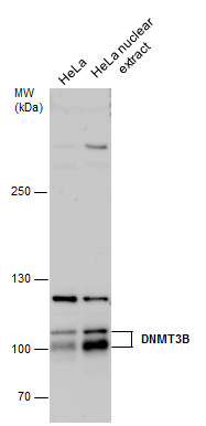
GTX129127 WB Image
DNMT3B antibody detects DNMT3B protein by western blot analysis. HeLa whole cell extracts and nuclear extracts (30 ug) were separated by 5% SDS-PAGE, and the membrane was blotted with DNMT3B antibody (GTX129127) diluted by 1:500.
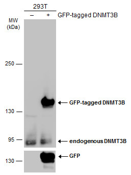
GTX129127 WB Image
Non-transfected (?) and transfected (+) 293T whole cell extracts (30 ug) were separated by 5% SDS-PAGE, and the membrane was blotted with DNMT3B antibody (GTX129127) diluted at 1:5000. The HRP-conjugated anti-rabbit IgG antibody (GTX213110-01) was used to detect the primary antibody, and the signal was developed with Trident ECL plus-Enhanced.
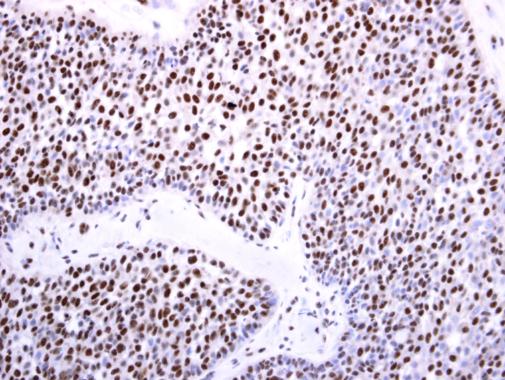
GTX129127 IHC-P Image
DNMT3B antibody detects DNMT3B protein at nucleus on human lung carcinoma by immunohistochemical analysis.
Sample: Paraffin-embedded human lung carcinoma.
DNMT3B antibody (GTX129127) diluted at 1:250.
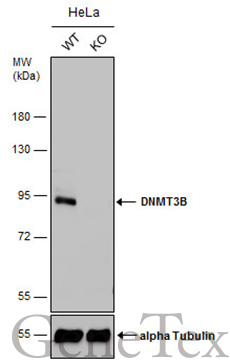
GTX129127 WB Image
Wild-type (WT) and DNMT3B knockout (KO) HeLa cell extracts (30 ug) were separated by 7.5% SDS-PAGE, and the membrane was blotted with DNMT3B antibody (GTX129127) diluted at 1:500. The HRP-conjugated anti-rabbit IgG antibody (GTX213110-01) was used to detect the primary antibody.