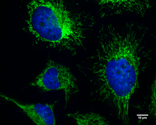
GTX129093 ICC/IF Image
FIP200 antibody detects FIP200 protein at cytoplasm by immunofluorescent analysis.
Sample: HeLa cells were fixed in 4% paraformaldehyde at RT for 10 min.
Green: FIP200 protein stained by FIP200 antibody (GTX129093) diluted at 1:200.
Blue: Hoechst 33342 staining.
Scale bar = 10 um.
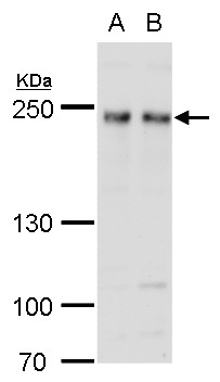
GTX129093 WB Image
FIP200 antibody detects FIP200 protein by western blot analysis.
A. 30 ug 293T whole cell lysate/extract
B. 30 ug A431 whole cell lysate/extract
5 % SDS-PAGE
FIP200 antibody (GTX129093) dilution: 1:1000
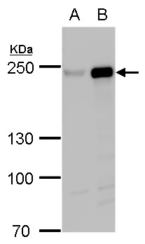
GTX129093 WB Image
FIP200 antibody detects FIP200 protein by western blot analysis.
A. 30 ug 293T whole cell lysate/extract
B. 30 ug whole cell lysate/extract of human FIP200-transfected 293T cells
5 % SDS-PAGE
FIP200 antibody (GTX129093) dilution: 1:5000