GTX128414 WB Image
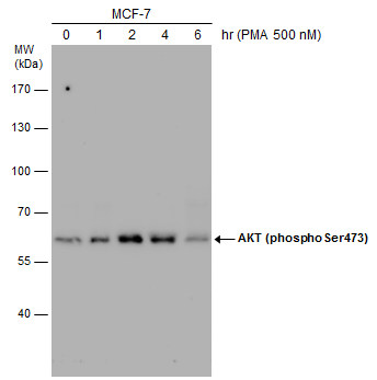
MCF-7 cells were untreated or treated with 500 nM PMA for 1, 2, 4 and 6 hrs. Various whole cell extracts (30 ug) were separated by 7.5% SDS-PAGE, and the membrane was blotted with AKT (phospho Ser473) antibody (GTX128414) diluted at 1:500. The HRP-conjugated anti-rabbit IgG antibody (GTX213110-01) was used to detect the primary antibody.
GTX128414 WB Image
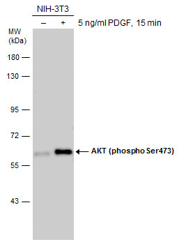
Untreated (?) and treated (+) NIH-3T3 whole cell extracts (30 ug) were separated by 7.5% SDS-PAGE, and the membrane was blotted with AKT (phospho Ser473) antibody (GTX128414) diluted at 1:500. The HRP-conjugated anti-rabbit IgG antibody (GTX213110-01) was used to detect the primary antibody.
GTX128414 WB Image
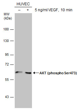
Untreated (?) and treated (+) HUVEC whole cell extracts (30 ug) were separated by 7.5% SDS-PAGE, and the membrane was blotted with AKT (phospho Ser473) antibody (GTX128414) diluted at 1:500. The HRP-conjugated anti-rabbit IgG antibody (GTX213110-01) was used to detect the primary antibody.
GTX128414 IP Image
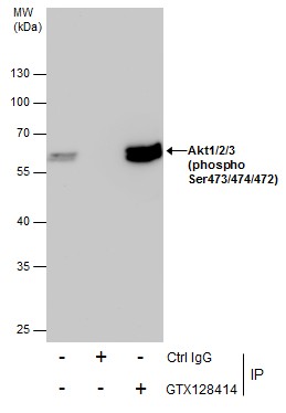
Immunoprecipitation of Akt1/2/3 (phospho Ser473/474/472) protein from 293T whole cell extracts using 5 ug of Akt1/2/3 (phospho Ser473/474/472) antibody (GTX128414).
Western blot analysis was performed using Akt1/2/3 (phospho Ser473/474/472) antibody (GTX128414).
EasyBlot anti-Rabbit IgG (GTX221666-01) was used as a secondary reagent.
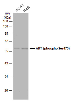
GTX128414 WB Image
Various whole cell extracts (30 ug) were separated by 10% SDS-PAGE, and the membrane was blotted with AKT (phospho Ser473) antibody (GTX128414) diluted at 1:500. The HRP-conjugated anti-rabbit IgG antibody (GTX213110-01) was used to detect the primary antibody, and the signal was developed with Trident ECL plus-Enhanced.