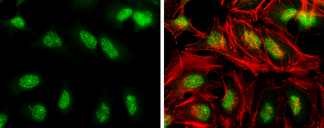
GTX128145 ICC/IF Image
ATR (phospho Thr1989) antibody detects ATR (phospho Thr1989) protein at nucleus by immunofluorescent analysis.Sample: HeLa cells were fixed in 4% paraformaldehyde at RT for 15 min.Green: ATR (phospho Thr1989) stained by ATR (phospho Thr1989) antibody (GTX128145) diluted at 1:500.Red: phalloidin, a cytoskeleton marker, diluted at 1:100.
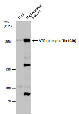
GTX128145 WB Image
Raji whole cell and nuclear extracts (30 ug) were separated by 5% SDS-PAGE, and the membrane was blotted with ATR (phospho Thr1989) antibody (GTX128145) diluted at 1:500. The HRP-conjugated anti-rabbit IgG antibody (GTX213110-01) was used to detect the primary antibody.
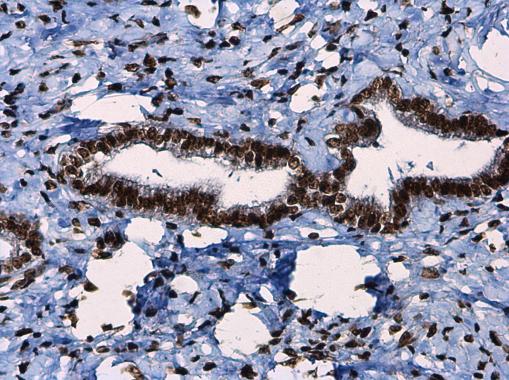
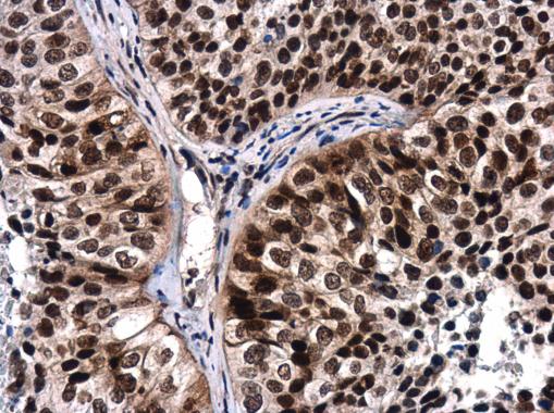
GTX128145 IHC-P Image
ATR (phospho Thr1989) antibody detects ATR (phospho Thr1989) protein at nucleus in human breast carcinoma by immunohistochemical analysis.
Sample: Paraffin-embedded human breast carcinoma.
ATR (phospho Thr1989) antibody (GTX128145) diluted at 1:250.
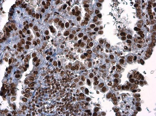
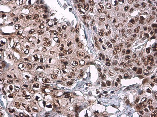
GTX128145 IHC-P Image
ATR (phospho Thr1989) antibody detects ATR (phospho Thr1989) protein at nucleus in human colon cancer by immunohistochemical analysis.
Sample: Paraffin-embedded human colon cancer.
ATR (phospho Thr1989) antibody (GTX128145) diluted at 1:250.

GTX128145 WB Image
Raji whole cell and nuclear extracts (30 ug) were separated by 5% SDS-PAGE, and the membranes were blotted with ATR (phospho Thr1989) antibody (GTX128145) diluted at 1:500 and competitor's antibody (CST#2790) diluted at 1:1000. The HRP-conjugated anti-rabbit IgG antibody (GTX213110-01) was used to detect the primary antibody.
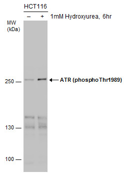
GTX128145 WB Image
Untreated (?) and treated (+) HCT-116 whole cell extracts (30 ug) were separated by 5% SDS-PAGE, and the membrane was blotted with ATR (phospho Thr1989) antibody (GTX128145) diluted at 1:1000.
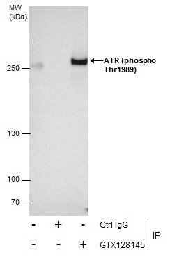
GTX128145 IP Image
Immunoprecipitation of ATR (phospho Thr1989) protein from HCT-116 whole cell extracts treated with Hydroxyurea for 6 hr using 5 ug of ATR (phospho Thr1989) antibody (GTX128145).
Western blot analysis was performed using ATR (phospho Thr1989) antibody (GTX128145).
EasyBlot anti-Rabbit IgG (GTX221666-01) was used as a secondary reagent.
GTX128145 IHC-P Image
ATR (phospho Thr1989) antibody detects ATR (phospho Thr1989) protein at nucleus in human lung by immunohistochemical analysis.
Sample: Paraffin-embedded human lung.
ATR (phospho Thr1989) antibody (GTX128145) diluted at 1:250.
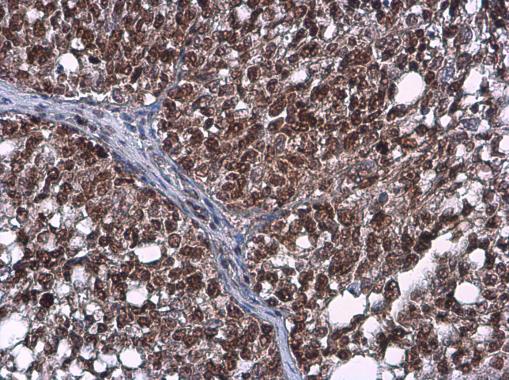
GTX128145 IHC-P Image
ATR (phospho Thr1989) antibody detects ATR (phospho Thr1989) protein at nucleus by immunohistochemical analysis.Sample: Paraffin-embedded human esophageal carcinoma.ATR (phospho Thr1989) stained by ATR (phospho Thr1989) antibody (GTX128145) diluted at 1:500.
GTX128145 IHC-P Image
ATR (phospho Thr1989) antibody detects ATR (phospho Thr1989) protein at nucleus by immunohistochemical analysis.Sample: Paraffin-embedded human ovarian cancer.ATR (phospho Thr1989) stained by ATR (phospho Thr1989) antibody (GTX128145) diluted at 1:500.
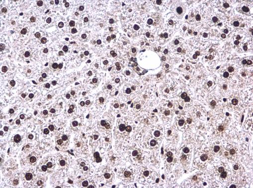
GTX128145 IHC-P Image
ATR (phospho Thr1989) antibody detects ATR (phospho Thr1989) protein at nucleus in mouse liver by immunohistochemical analysis.
Sample: Paraffin-embedded mouse liver.
ATR (phospho Thr1989) antibody (GTX128145) diluted at 1:200.
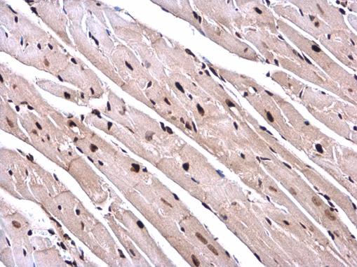
GTX128145 IHC-P Image
ATR (phospho Thr1989) antibody detects ATR (phospho Thr1989) protein at nucleus in mouse heart by immunohistochemical analysis.
Sample: Paraffin-embedded mouse heart.
ATR (phospho Thr1989) antibody (GTX128145) diluted at 1:200.
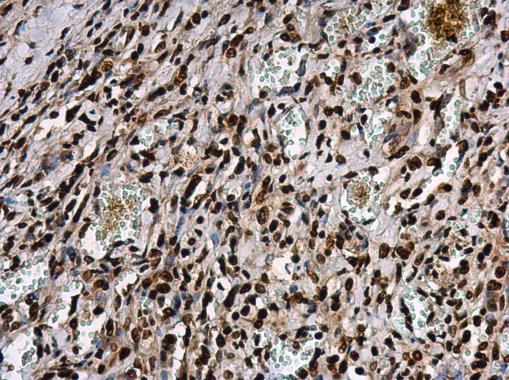
GTX128145 IHC-P Image
ATR (phospho Thr1989) antibody detects ATR protein at nucleus in human lung cancer by immunohistochemical analysis.
Sample: Paraffin-embedded human lung cancer.
ATR (phospho Thr1989) antibody (GTX128145) diluted at 1:250.