GTX127345 IHC-Fr Image
N-Cadherin antibody detects N-Cadherin protein expression by immunohistochemical analysis.
Sample: Frozen sectioned E13.5 Rat brain.
Green: N-Cadherin protein stained by N-Cadherin antibody (GTX127345) diluted at 1:250.
Red: beta Tubulin 3/ TUJ1, a mature neuron marker, stained by beta Tubulin 3/ TUJ1 antibody [GT11710] (GTX631836) diluted at 1:500.
Blue: Fluoroshield with DAPI (GTX30920).
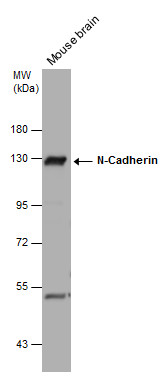
GTX127345 WB Image
Mouse tissue extract (50 ug) was separated by 7.5% SDS-PAGE, and the membrane was blotted with N-Cadherin antibody (GTX127345) diluted at 1:1000. The HRP-conjugated anti-rabbit IgG antibody (GTX213110-01) was used to detect the primary antibody.
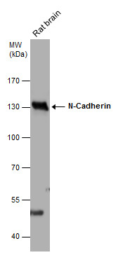
GTX127345 WB Image
Rat tissue extract (50 ug) was separated by 7.5% SDS-PAGE, and the membrane was blotted with N-Cadherin antibody (GTX127345) diluted at 1:1000. The HRP-conjugated anti-rabbit IgG antibody (GTX213110-01) was used to detect the primary antibody.
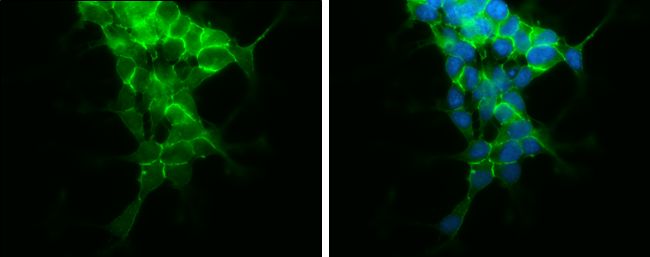
GTX127345 ICC/IF Image
N-Cadherin antibody detects N-Cadherin protein at cell membrane by immunofluorescent analysis.
Sample: SH-SY5Y cells were fixed in 4% paraformaldehyde at RT for 15 min.
Green: N-Cadherin protein stained by N-Cadherin antibody (GTX127345) diluted at 1:500.
Blue: Hoechst 33342 staining.
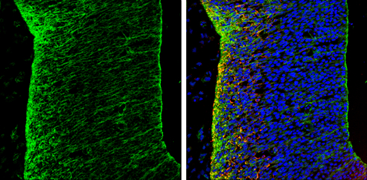
GTX127345 IHC-Fr Image
N-Cadherin antibody detects N-Cadherin protein on embryonic mouse brain by immunohistochemical analysis. Sample: Frozen section of embryonic mouse brain (mE18.5). N-Cadherin antibody (GTX127345) diluted at 1:500.
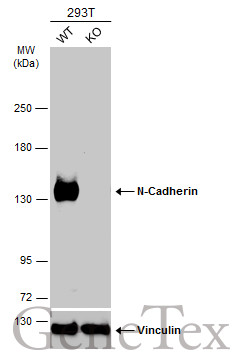
GTX127345 WB Image
Wild-type (WT) and N-Cadherin knockout (KO) 293T cell extracts (30 ug) were separated by 5% SDS-PAGE, and the membrane was blotted with N-Cadherin antibody (GTX127345) diluted at 1:500. The HRP-conjugated anti-rabbit IgG antibody (GTX213110-01) was used to detect the primary antibody.
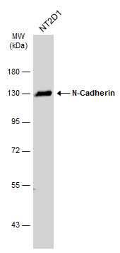
GTX127345 WB Image
Whole cell extract (30 ug) was separated by 7.5% SDS-PAGE, and the membrane was blotted with N-Cadherin antibody (GTX127345) diluted at 1:1000. The HRP-conjugated anti-rabbit IgG antibody (GTX213110-01) was used to detect the primary antibody.
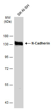
GTX127345 WB Image
Whole cell extract (30 ug) was separated by 7.5% SDS-PAGE, and the membrane was blotted with N-Cadherin antibody (GTX127345) diluted at 1:1000. The HRP-conjugated anti-rabbit IgG antibody (GTX213110-01) was used to detect the primary antibody.
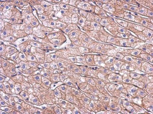
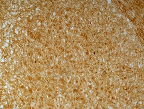
GTX127345 IHC-P Image
Immunohistochemical analysis of paraffin-embedded human hepatoma, using N-Cadherin antibody(GTX127345) antibody at 1:500 dilution.