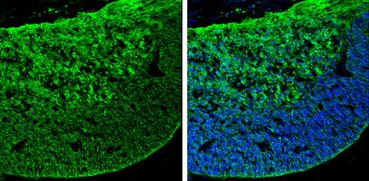
GTX125988 IHC-Fr Image
GABA antibody detects GABA by immunohistochemical analysis.
Sample: Frozen sectioned E13.5 Rat brain.
Green: GABA stained by GABA antibody (GTX125988) diluted at 1:250.
Blue: Fluoroshield with DAPI (GTX30920).
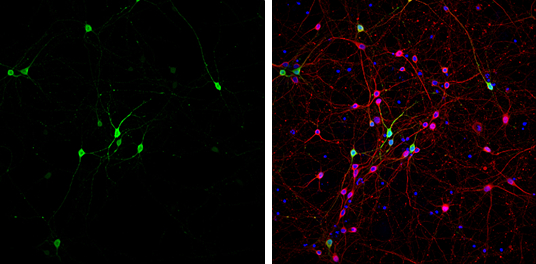
GTX125988 ICC/IF Image
GABA antibody detects GABA at GABAergic neurons by immunofluorescent analysis.
Sample: DIV9 rat E18 primary cortical neurons were fixed in 4% paraformaldehyde at RT for 15 min.
Green: GABA stained by GABA antibody (GTX125988) diluted at 1:500.
Red: beta Tubulin 3/ Tuj1, stained by beta Tubulin 3/ Tuj1 antibody [GT11710] (GTX631836) diluted at 1:500.
Blue: Fluoroshield with DAPI (GTX30920).
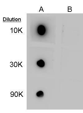
GTX125988 Dot Image
Dot blot analysis of 100ng of GABA-conjugated BSA, using GABA Antibody (GTX125988) at 1:10000, 1:30000, and 1:90000. A: GABA-conjugated BSA. B: Normal BSA
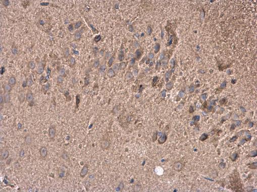
GTX125988 IHC-P Image
GABA antibody detects GABA protein at cytoplasm in rat brain by immunohistochemical analysis.
Sample: Paraffin-embedded rat brain.
GABA antibody (GTX125988) diluted at 1:400.