GTX125891 IP Image
Paxillin antibody immunoprecipitates Paxillin protein in IP experiments.
IP samples: HeLa whole cell extract
A. Control with 2 ug of preimmune Rabbit IgG
B. Immunoprecipitation of Paxillin protein by 2 ug Paxillin antibody (GTX125891)
7.5 % SDS-PAGE
The immunoprecipitated Paxillin protein was detected by Paxillin antibody (GTX125891) diluted at 1:10000.
[EasyBlot anti-rabbit IgG (GTX221666-01) was used as a secondary reagent]
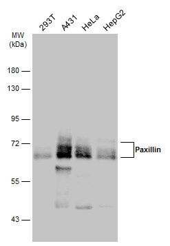
GTX125891 WB Image
Various whole cell extracts (30 ug) were separated by 7.5% SDS-PAGE, and the membrane was blotted with Paxillin antibody (GTX125891) diluted at 1:5000. The HRP-conjugated anti-rabbit IgG antibody (GTX213110-01) was used to detect the primary antibody.
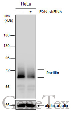
GTX125891 WB Image
Non-transfected (?) and transfected (+) HeLa whole cell extracts (30 ug) were separated by 7.5% SDS-PAGE, and the membrane was blotted with Paxillin antibody (GTX125891) diluted at 1:6000. The HRP-conjugated anti-rabbit IgG antibody (GTX213110-01) was used to detect the primary antibody.
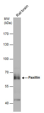
GTX125891 WB Image
Rat tissue extract (50 ug) was separated by 7.5% SDS-PAGE, and the membrane was blotted with Paxillin antibody (GTX125891) diluted at 1:1000. The HRP-conjugated anti-rabbit IgG antibody (GTX213110-01) was used to detect the primary antibody.
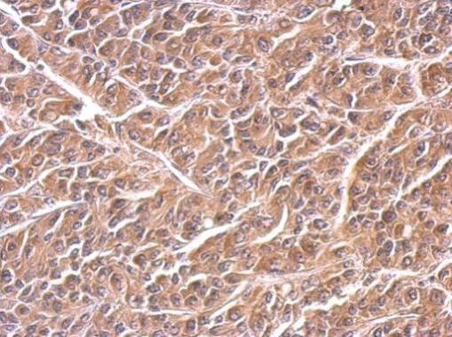
GTX125891 IHC-P Image
Paxillin antibody detects PXN protein at cytosol on AGS xenograft by immunohistochemical analysis.
Sample: Paraffin-embedded AGS xenograft.
Paxillin antibody (GTX125891) dilution: 1:500.
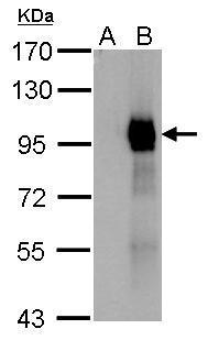
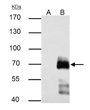
GTX125891 WB Image
Paxillin antibody detects Paxillin protein by western blot analysis.
A. 1 ug 293T whole cell extract
B. 1 ug whole cell extract of GFP-human Paxillin-transfected 293T cells
7.5% SDS-PAGE
Paxillin antibody (GTX125891) dilution: 1:10000
The HRP-conjugated anti-rabbit IgG antibody (GTX213110-01) was used to detect the primary antibody.
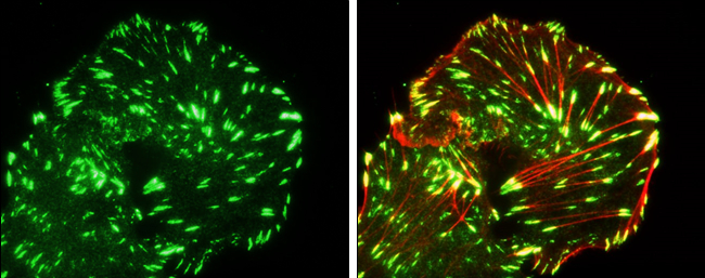
GTX125891 ICC/IF Image
Paxillin antibody detects Paxillin protein at cytoskeleton by immunofluorescent analysis.
Sample: MDA-MB-231 cells were fixed in 4% paraformaldehyde at RT for 15 min.
Green: Paxillin protein stained by Paxillin antibody (GTX125891) diluted at 1:100.
Red: phalloidin staining.
Courtesy of a researcher who prefers to be anonymous.