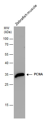
GTX124496 WB Image
PCNA antibody detects PCNA protein by western blot analysis. Zebrafish tissue extracts (30 ug) was separated by 12% SDS-PAGE, and the membrane was blotted with PCNA antibody (GTX124496) at a dilution of 1:1000. The HRP-conjugated anti-rabbit IgG antibody (GTX213110-01) was used to detect the primary antibody.
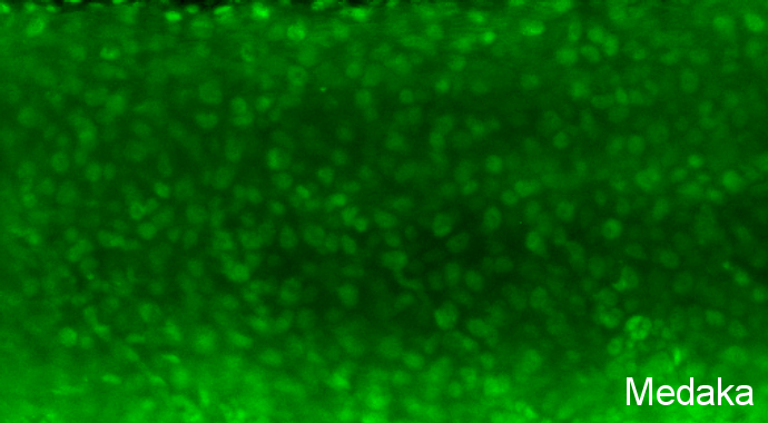
GTX124496 IHC-Wm Image
Pcna antibody detects Pcna protein on Medaka by whole mount immunohistochemical analysis.
Sample: 7 days-post-fertilization medaka embryo.
Pcna antibody (GTX124496) dilution: 1:100.
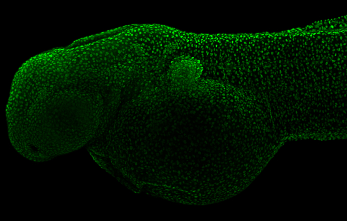
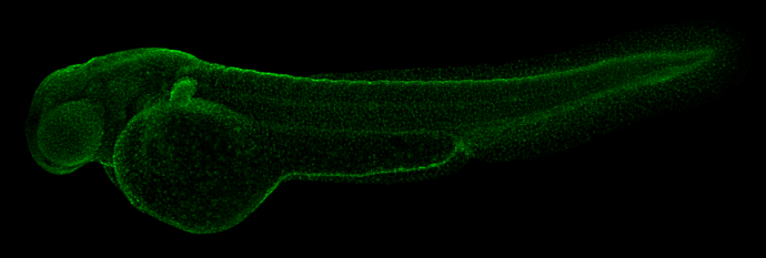
GTX124496 IHC-Wm Image
Pcna antibody detects Pcna protein on whole mount zebrafish by immunohistochemical analysis.
Sample: 2 day-post-fertilization zebrafish embryo.
Pcna antibody (GTX124496) dilution: 1:100.
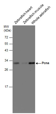
GTX124496 WB Image
Various tissue extracts (30 ug) were separated by 12% SDS-PAGE, and the membrane was blotted with Pcna antibody (GTX124496) diluted at 1:500. The HRP-conjugated anti-rabbit IgG antibody (GTX213110-01) was used to detect the primary antibody.
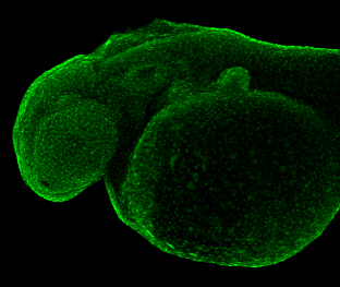
GTX124496 IHC-Wm Image
Pcna antibody detects Pcna protein on zebrafish by whole mount immunohistochemical analysis.
Sample: Paraformaldehyde-fixed 2 day-post-fertilization zebrafish embryo.
Pcna antibody (GTX124496) dilution: 1:50.
GTX124496 IHC-Wm Image
Pcna antibody detects Pcna protein on zebrafish by whole mount immunohistochemical analysis.
Sample: Paraformaldehyde-fixed 2 day-post-fertilization zebrafish embryo.
Pcna antibody (GTX124496) dilution: 1:50.

GTX124496 IHC-P Image
Immunohistochemical analysis of paraffin-embedded zebrafish kidney, using pcna (GTX124496) antibody at 1:300 dilution.