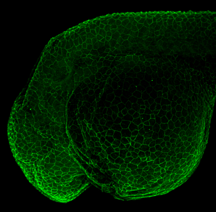
GTX124481 IHC-Wm Image
Ctnnb1 antibody detects Ctnnb1 protein on zebrafish by whole mount immunohistochemical analysis.
Sample: 1 day-post-fertilization zebrafish embryo.
Ctnnb1 antibody (GTX124481) dilution: 1:100.
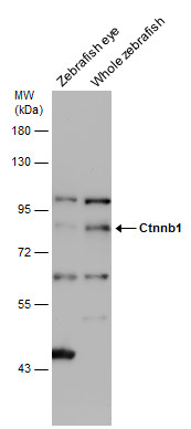
GTX124481 WB Image
Various tissue extracts (30 ug) were separated by 7.5% SDS-PAGE, and the membrane was blotted with Ctnnb1 antibody (GTX124481) diluted at 1:500.
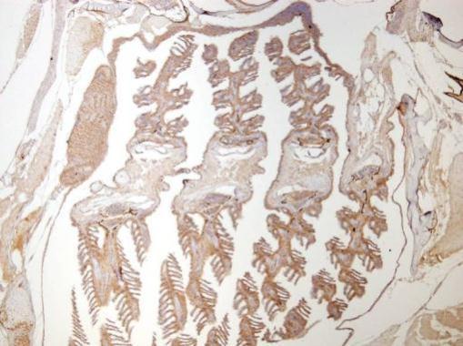
GTX124481 IHC-P Image
Immunohistochemical analysis of paraffin-embedded zebrafish tissue, using ctnnb1 (GTX124481) antibody at 1:300 dilution.
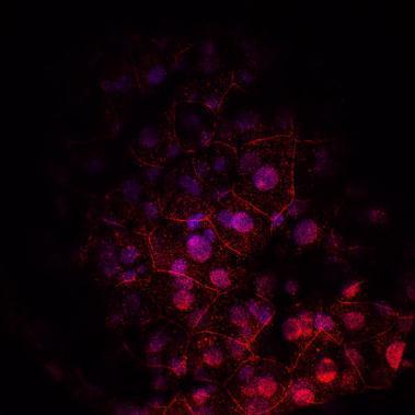
GTX124481 IHC Image
Immunohistochemical analysis of agarose-embedded zebrafish embryo, using ctnnb1 (GTX124481) antibody at 1:100 dilution. (This image was provided courtesy of the Schilling Lab at UC, Irvine.)
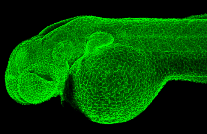
GTX124481 IHC-Wm Image
Ctnnb1 antibody detects Ctnnb1 protein on zebrafish by whole mount immunohistochemical analysis.
Sample: 2 days-post-fertilization zebrafish embryo.
Ctnnb1 antibody (GTX124481) dilution: 1:100.
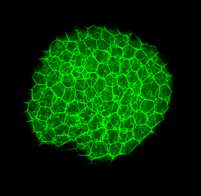
GTX124481 IHC-Wm Image
Ctnnb1 antibody detects Ctnnb1 protein on zebrafish by whole mount immunohistochemical analysis.
Sample: 2 days-post-fertilization zebrafish embryo.
Ctnnb1 antibody (GTX124481) dilution: 1:100.