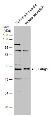
GTX124352 WB Image
Various tissue extracts (30 ug) were separated by 10% SDS-PAGE, and the membrane was blotted with Tubg1 antibody (GTX124352) diluted at 1:1000. The HRP-conjugated anti-rabbit IgG antibody (GTX213110-01) was used to detect the primary antibody.
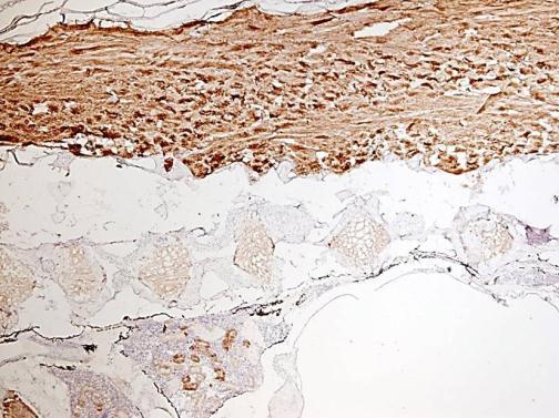
GTX124352 IHC Image
Immunohistochemical analysis of agarose-embedded zebrafish embryo, using tubgl (GTX124352) antibody at 1:100 dilution. (This image was provided courtesy of the Schilling Lab at UC, Irvine.)
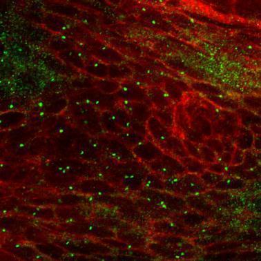
GTX124352 IHC-P Image
Immunohistochemical analysis of paraffin-embedded sections of zebrafish neurons, using tubgl (GTX124352) antibody at 1:300 dilution.
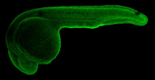
GTX124352 IHC-Wm Image
Tubg1 antibody detects Tubg1 protein on whole mount zebrafish by immunohistochemical analysis. Sample: 1 day-post-fertilization zebrafish embryo.Tubg1 antibody (GTX124352) dilution: 1:100.