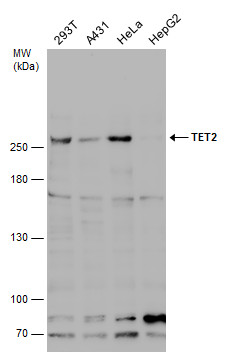
GTX124204 WB Image
Various whole cell extracts (30 ug) were separated by 5% SDS-PAGE, and the membrane was blotted with TET2 antibody [N2], N-term (GTX124204) diluted at 1:500. The HRP-conjugated anti-rabbit IgG antibody (GTX213110-01) was used to detect the primary antibody.
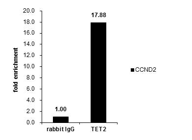
GTX124204 ChIP assay Image
Cross-linked ChIP was performed with U2OS chromatin extract and 5 ug of either control rabbit IgG or anti-TET2 antibody. The precipitated DNA was detected by PCR with primer set targeting to CCND2.
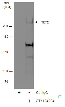
GTX124204 IP Image
Immunoprecipitation of TET2 protein from 293T whole cell extracts using 5 ug of TET2 antibody [N2], N-term (GTX124204).
Western blot analysis was performed using TET2 antibody [N2], N-term (GTX124204).
EasyBlot HRP-conjugated anti rabbit IgG antibody (GTX221666-01) was used to detect the primary antibody.
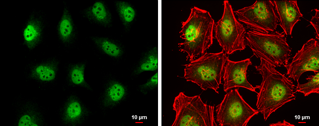
GTX124204 ICC/IF Image
TET2 antibody [N2], N-term detects TET2 protein at nucleus by immunofluorescent analysis.
Sample: HeLa cells were fixed in 4% paraformaldehyde at RT for 15 min.
Green: TET2 protein stained by TET2 antibody [N2], N-term (GTX124204) diluted at 1:500.
Red: phalloidin, a cytoskeleton marker, diluted at 1:200.
Scale bar = 10 um.
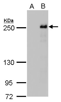
GTX124204 WB Image
TET2 antibody detects TET2 protein by western blot analysis.
A. 30 ug 293T whole cell lysate/extract
B. 30 ug whole cell lysate/extract of DDDDK-human TET2-transfected 293T cells
5% SDS-PAGE
TET2 antibody (GTX124204) dilution: 1:5000
The HRP-conjugated anti-rabbit IgG antibody (GTX213110-01) was used to detect the primary antibody.