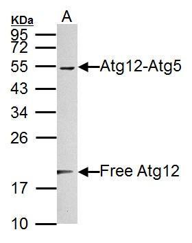
GTX124181 WB Image
Sample (30 ug of whole cell lysate)
A: NIH-3T3
12% SDS PAGE
GTX124181 diluted at 1:1000
The HRP-conjugated anti-rabbit IgG antibody (GTX213110-01) was used to detect the primary antibody.
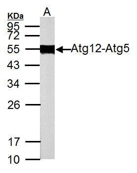
GTX124181 WB Image
Sample (50 ug of whole cell lysate)
A: Rat brain
12% SDS PAGE
GTX124181 diluted at 1:1000
The HRP-conjugated anti-rabbit IgG antibody (GTX213110-01) was used to detect the primary antibody.
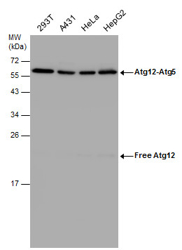
GTX124181 WB Image
Various whole cell extracts (30 ug) were separated by 12% SDS-PAGE, and the membrane was blotted with ATG12 antibody (GTX124181) diluted at 1:1000.
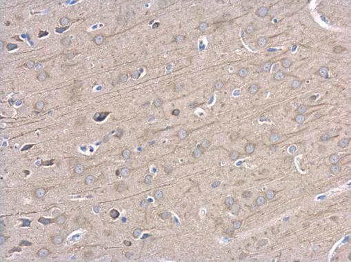
GTX124181 IHC-P Image
ATG12 antibody detects ATG12 protein at cytoplasm in rat brain by immunohistochemical analysis.
Sample: Paraffin-embedded rat brain.
ATG12 antibody (GTX124181) diluted at 1:500.
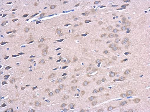
GTX124181 IHC-P Image
ATG12 antibody detects ATG12 protein at cytoplasm in rat brain by immunohistochemical analysis.
Sample: Paraffin-embedded rat brain.
ATG12 antibody (GTX124181) diluted at 1:500.
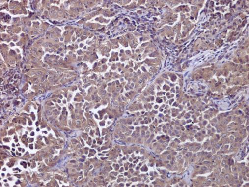
GTX124181 IHC-P Image
ATG12 antibody detects ATG12 protein at cytoplasm in human lung cancer by immunohistochemical analysis.
Sample: Paraffin-embedded human lung cancer.
ATG12 antibody (GTX124181) diluted at 1:500.
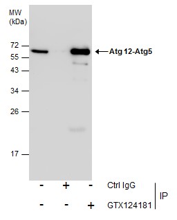
GTX124181 IP Image
Immunoprecipitation of Atg 12-Atg5 protein from 293T whole cell extracts using 5 ug of ATG12 antibody (GTX124181).
Western blot analysis was performed using ATG12 antibody (GTX124181).
EasyBlot anti-Rabbit IgG (GTX221666-01) was used as a secondary reagent.
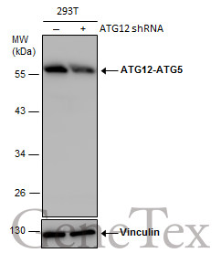
GTX124181 WB Image
Non-transfected (?) and transfected (+) 293T whole cell extracts (30 ug) were separated by 10% SDS-PAGE, and the membrane was blotted with ATG12 antibody (GTX124181) diluted at 1:500. The HRP-conjugated anti-rabbit IgG antibody (GTX213110-01) was used to detect the primary antibody.