GTX122648 WB Image
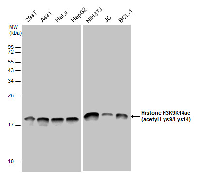
Histone H3K9K14ac (acetyl Lys9/Lys14) antibody detects Histone H3K9K14ac (acetyl Lys9/Lys14) protein by western blot analysis. Various whole cell extracts (30 ug) were separated by 12% SDS-PAGE, and the membrane was blotted with Histone H3K9K14ac (acetyl Lys9/Lys14) antibody (GTX122648) diluted at a dilution of 1:10000. The HRP-conjugated anti-rabbit IgG antibody (GTX213110-01) was used to detect the primary antibody.

GTX122648 ICC/IF Image
Histone H3K9K14ac (acetyl Lys9/Lys14) antibody detects Histone H3K9K14ac (acetyl Lys9/Lys14) protein at nucleus by immunofluorescent analysis.
Sample: HeLa cells were fixed in 4% paraformaldehyde at RT for 15 min.
Green: Histone H3K9K14ac (acetyl Lys9/Lys14) protein stained by Histone H3K9K14ac (acetyl Lys9/Lys14) antibody (GTX122648) diluted at 1:1000.
Red: alpha Tubulin, a cytoskeleton marker, stained by alpha Tubulin antibody [GT114] (GTX628802) diluted at 1:1000.
Blue: Hoechst 33342 staining.
GTX122648 WB Image
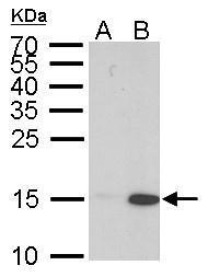
Histone H3 (acetyl Lys 9, Lys14) antibody detects Histone H3 (acetyl Lys 9, Lys14) protein by western blot analysis.
A. 30 ug HeLa whole cell lysate/extract (untreated)
B. 30 ug HeLa whole cell lysate/extract (0.4 uM Trichostatin treatment for 18 hr)
15% SDS-PAGE
Histone H3 (acetyl Lys 9, Lys14) antibody (GTX122648) dilution: 1:10000
The HRP-conjugated anti-rabbit IgG antibody (GTX213110-01) was used to detect the primary antibody.
GTX122648 Dot Image
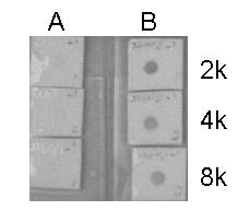
Dotblot analysis of anti-Histone H3 (acetyl Lys 9, Lys14) antibody with peptide samples.
Peptide samples (0.1 ug) were spotted onto positively charged nylon membrane and blotted with Histone H3 (acetyl Lys 9, Lys14) antibody (GTX122648) at different dilution as indicated.
A: Peptide samples of Histone H3.1
B: Peptide samples of Histone H3 (acetyl Lys 9, Lys14)