
GTX121937 IHC Image
Staining of Human normal esophagus tissue sections using anti-Akt1/2/3 antibody (GTX121937).
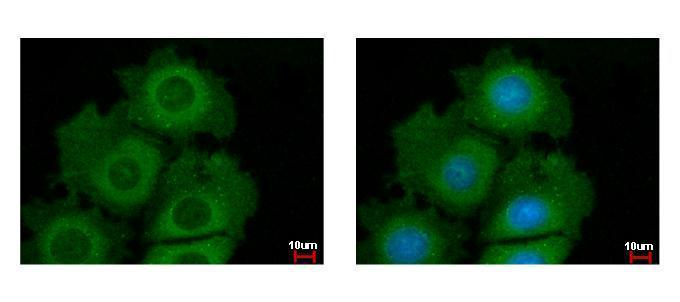
GTX121937 ICC/IF Image
Akt1/2/3 antibody detects Akt1/2/3 protein at cytoplasm by immunofluorescent analysis.
Sample: MCF-7 cells were fixed in ice-cold MeOH for 5 min.
Green: Akt1 protein stained by Akt1/2/3 antibody (GTX121937) diluted at 1:500.
Blue: Hoechst 33342 staining.

GTX121937 WB Image
Sample (30 ug of whole cell lysate)
A: JC
B: BCL-1
7.5% SDS PAGE
GTX121937 diluted at 1:5000
The HRP-conjugated anti-rabbit IgG antibody (GTX213110-01) was used to detect the primary antibody.
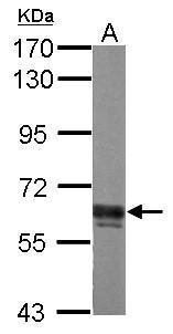
GTX121937 WB Image
Sample (30 ug of whole cell lysate)
A: PC-12
7.5% SDS PAGE
GTX121937 diluted at 1:3000
The HRP-conjugated anti-rabbit IgG antibody (GTX213110-01) was used to detect the primary antibody.

GTX121937 WB Image
Non-transfected (?) and transfected (+) HeLa whole cell extracts (30 ug) were separated by 7.5% SDS-PAGE, and the membrane was blotted with AKT antibody [N3C2], Internal (GTX121937) diluted at 1:5000. The HRP-conjugated anti-rabbit IgG antibody (GTX213110-01) was used to detect the primary antibody.

GTX121937 IP Image
Immunoprecipitation of Akt1/2/3 protein from 293T whole cell extracts using 5 ug of Akt1/2/3 antibody [N3C2], Internal (GTX121937).
Western blot analysis was performed using Akt1/2/3 antibody [N3C2], Internal (GTX121937).
EasyBlot anti-Rabbit IgG (GTX221666-01) was used as a secondary reagent.
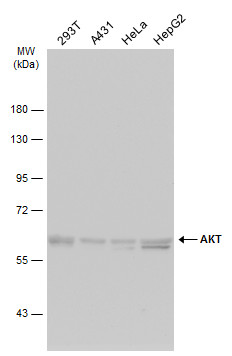
GTX121937 WB Image
Various whole cell extracts (30 ug) were separated by 7.5% SDS-PAGE, and the membrane was blotted with AKT antibody [N3C2], Internal (GTX121937) diluted at 1:1000. The HRP-conjugated anti-rabbit IgG antibody (GTX213110-01) was used to detect the primary antibody, and the signal was developed with Trident ECL plus-Enhanced.