
GTX121936 WB Image
Akt1/2/3 antibody detects Akt1/2/3 protein by Western blot analysis.
A. 30 ug 293T whole cell lysate/extract
B. 30 whole cell lysate/extract of HA-human Akt-transfected 293T cells
7.5 % SDS-PAGE
Akt1/2/3 antibody (GTX121936) dilution: 1:5000

GTX121936 IHC Image
Staining of Human esophageal cancer tissue sections using anti-Akt1/2/3 antibody (GTX121936).
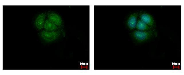
GTX121936 ICC/IF Image
Akt1/2/3 antibody detects Akt1/2/3 protein at cytoplasm and nucleus by immunofluorescent analysis.
Sample: HeLa cells were fixed in ice-cold MeOH for 5 min.
Green: Akt1 protein stained by Akt1/2/3 antibody (GTX121936) diluted at 1:500.
Blue: Hoechst 33342 staining.