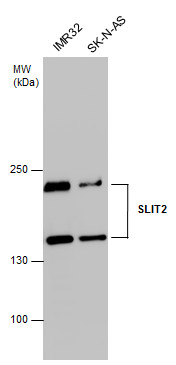
GTX118220 WB Image
SLIT2 antibody detects SLIT2 protein by western blot analysis. Various whole cell extracts (30 ug) were separated by 5% SDS-PAGE, and the membrane was blotted with SLIT2 antibody (GTX118220) diluted at 1:1000. The HRP-conjugated anti-rabbit IgG antibody (GTX213110-01) was used to detect the primary antibody.
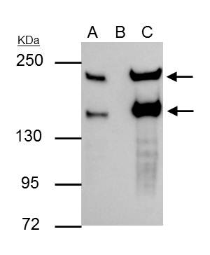
GTX118220 IP Image
SLIT2 antibody immunoprecipitates SLIT2 protein in IP experiments. IP Sample: Raji whole cell lysate/extract A : 30 ug whole cell lysate/extract of SLIT2 protein expressing Raji cells B : Control with 3 ug of pre-immune rabbit IgG C : Immunoprecipitation of SLIT2 by 3 ug of SLIT2 antibody (GTX118220) 5% SDS-PAGE The immunoprecipitated SLIT2 protein was detected by SLIT2 antibody (GTX118220) diluted at 1 : 1000. EasyBlot anti-rabbit IgG (HRP) (GTX221666-01) was used as a secondary reagent.
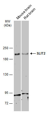
GTX118220 WB Image
Various tissue extracts (30 ug) were separated by 5% SDS-PAGE, and the membrane was blotted with SLIT2 antibody (GTX118220) diluted at 1:500. The HRP-conjugated anti-rabbit IgG antibody (GTX213110-01) was used to detect the primary antibody.
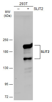
GTX118220 WB Image
Non-transfected (?) and transfected (+) 293T whole cell extracts (30 ug) were separated by 5% SDS-PAGE, and the membrane was blotted with SLIT2 antibody (GTX118220) diluted at 1:4000. The HRP-conjugated anti-rabbit IgG antibody (GTX213110-01) was used to detect the primary antibody.
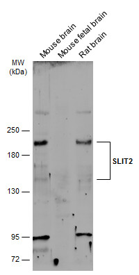
GTX118220 WB Image
Various tissue extracts (50 ug) were separated by 5% SDS-PAGE, and the membrane was blotted with SLIT2 antibody (GTX118220) diluted at 1:500. The HRP-conjugated anti-rabbit IgG antibody (GTX213110-01) was used to detect the primary antibody.
GTX118220 IHC-P Image
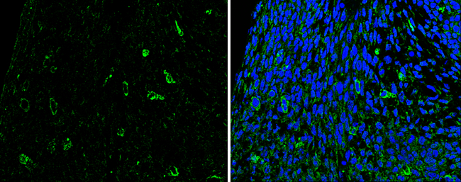
SLIT2 antibody detects SLIT2 protein at cytoplasm in mouse fetal brain by immunohistochemical analysis.
Sample: Paraffin-embedded mouse fetal brain.
Green: SLIT2 antibody (GTX118220) diluted at 1:200. The signal was developed using goat anti-rabbit IgG antibody (Dylight488) (GTX213110-04).
Blue: Nuclear staining with Hoechst 33342.
GTX118220 IHC-P Image
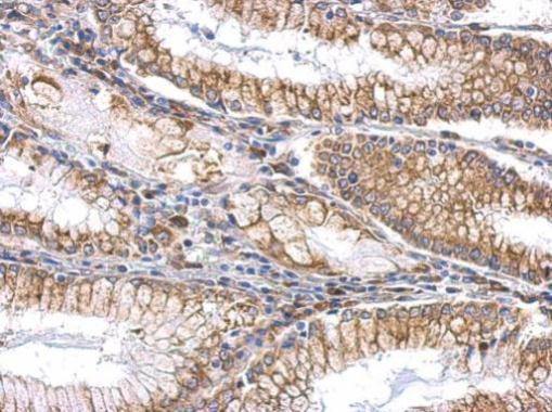
SLIT2 antibody detects SLIT2 protein at cytosol and membrane on gastric carcinoma by immunohistochemical analysis.
Sample: Paraffin-embedded human gastric carcinoma.
SLIT2 antibody (GTX118220) dilution: 1:500.
GTX118220 IHC-P Image
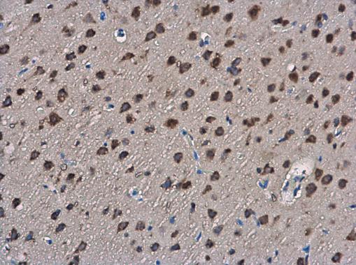
SLIT2 antibody detects SLIT2 protein at cytoplasm in rat brain by immunohistochemical analysis.
Sample: Paraffin-embedded rat brain.
SLIT2 antibody (GTX118220) diluted at 1:400.