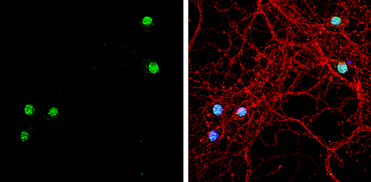
GTX117615 ICC/IF Image
TBR1 antibody [N2C1], Internal detects TBR1 protein by immunofluorescent analysis.Sample: DIV9 rat E18 primary hippocampal neuron cells were fixed in 4% paraformaldehyde at RT for 15 min.Green: TBR1 stained by TBR1 antibody [N2C1], Internal (GTX117615) diluted at 1:500.Red: beta Tubulin 3/ Tuj1, stained by beta Tubulin 3/ Tuj1 antibody [GT1338] (GTX631831) diluted at 1:500.Blue: Fluoroshield with DAPI (GTX30920).
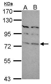
GTX117615 WB Image
Sample (30 ug of whole cell lysate)
A: Neuro2A
B: GL261
7.5% SDS PAGE
GTX117615 diluted at 1:1000
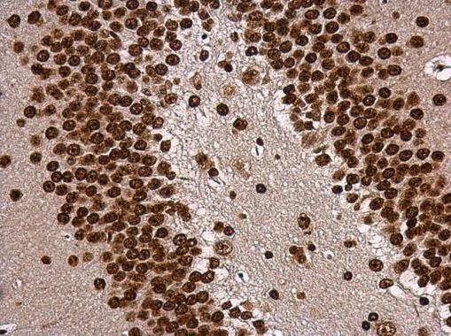
GTX117615 IHC-P Image
TBR1 antibody [N2C1], Internal detects TBR1 protein at nucleus in mouse brain by immunohistochemical analysis.
Sample: Paraffin-embedded mouse brain.
TBR1 antibody [N2C1], Internal (GTX117615) diluted at 1:500.