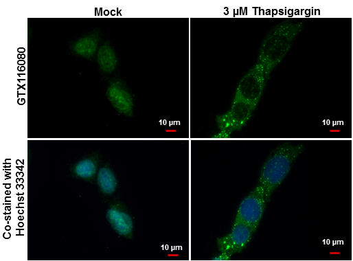
GTX116080 ICC/IF Image
LC3B antibody [N1C3] detects LC3B protein at autophagosome by immunofluorescent analysis.
Samples: Hep G2 cells mock (left) and treated with 3 uM Thapsigargin for 12 hrs (right) were fixed in ice-cold MeOH for 10 min and permeabilized with ice-cold acetone for 1 min.
Green: LC3B protein stained by LC3B antibody [N1C3] (GTX116080) diluted at 1:500.
Blue: Hoechst 33342 staining.
Scale bar = 10 um.
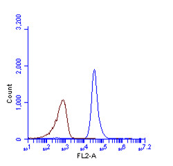
GTX116080 FACS Image
LC3B antibody [N1C3] (GTX116080) detects LC3B protein by flow cytometry analysis.
Sample: HeLa cell fixed in 4% paraformaldehyde at 4oC for 5 min.
Brown: Unlabelled sample was also used as a control.
Blue: LC3B antibody [N1C3] (GTX116080) dilution: 1:100.
Acquisition of >20,000 events were collected using Argon ion laser (488nm) and 525/30 bandpass filter.
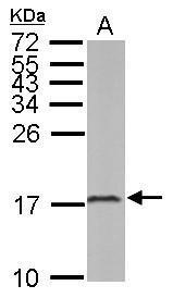
GTX116080 WB Image
Sample (50 ug of whole cell lysate)
A: mouse brain
15% SDS PAGE
GTX116080 diluted at 1:1000
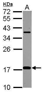
GTX116080 WB Image
Sample (50 ug of whole cell lysate)
A: Rat brain
12% SDS PAGE
GTX116080 diluted at 1:1000
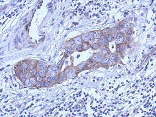
GTX116080 IHC-P Image
Immunohistochemical analysis of paraffin-embedded human Breast carcinoma, using LC3B(GTX116080) antibody at 1:500 dilution.
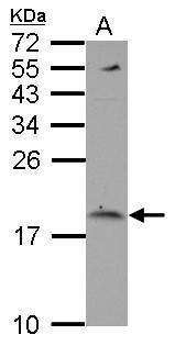
GTX116080 WB Image
Sample (30 ug of whole cell lysate)
A: U87-MG
15% SDS PAGE
GTX116080 diluted at 1:1000