GTX116041 WB Image
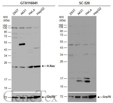
Various whole cell extracts (30 ug) were separated by 12% SDS-PAGE, and the membranes were blotted with H-Ras antibody (GTX116041) diluted at 1:500 and competitor's antibody (SC-520) diluted by 1:200. The HRP-conjugated anti-rabbit IgG antibody (GTX213110-01) was used to detect the primary antibody.
GTX116041 WB Image
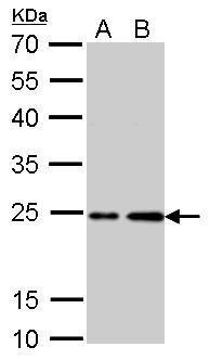
H-Ras antibody detects HRAS protein by western blot analysis.
A. 30 ug PC-12 whole cell lysate/extract
B. 30 ug Rat2 whole cell lysate/extract
12% SDS-PAGE
H-Ras antibody (GTX116041) dilution: 1:1000
The HRP-conjugated anti-rabbit IgG antibody (GTX213110-01) was used to detect the primary antibody.
GTX116041 ICC/IF Image
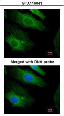
Immunofluorescence analysis of paraformaldehyde-fixed HeLa, using H-Ras (GTX116041) antibody at 1:200 dilution.
GTX116041 IHC-P Image
H-Ras antibody detects H-Ras protein at membrane on mouse prostate by immunohistochemical analysis.
Sample: Paraffin-embedded mouse prostate.
H-Ras antibody (GTX116041) dilution: 1:500.
GTX116041 IHC-P Image
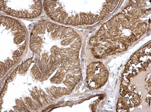
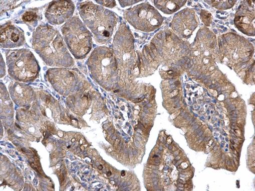
H-Ras antibody detects H-Ras protein at membrane on mouse intestine by immunohistochemical analysis.
Sample: Paraffin-embedded mouse intestine.
H-Ras antibody (GTX116041) dilution: 1:500.
GTX116041 WB Image
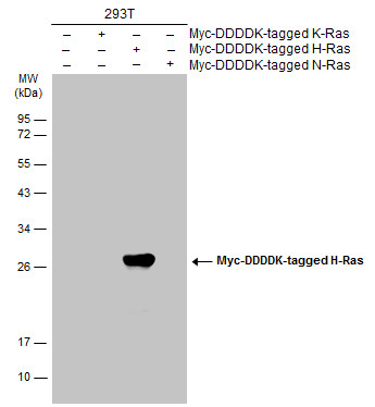
Non-transfected (?) and transfected (+) 293T whole cell extracts (30 ug) were separated by 12% SDS-PAGE, and the membrane was blotted with Myc-DDDDK-tagged H-Ras antibody (GTX116041) diluted at 1:5000. The HRP-conjugated anti-rabbit IgG antibody (GTX213110-01) was used to detect the primary antibody.
GTX116041 WB Image
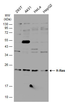
Various whole cell extracts (30 ug) were separated by 12% SDS-PAGE, and the membrane was blotted with H-Ras antibody (GTX116041) diluted at 1:500. The HRP-conjugated anti-rabbit IgG antibody (GTX213110-01) was used to detect the primary antibody.