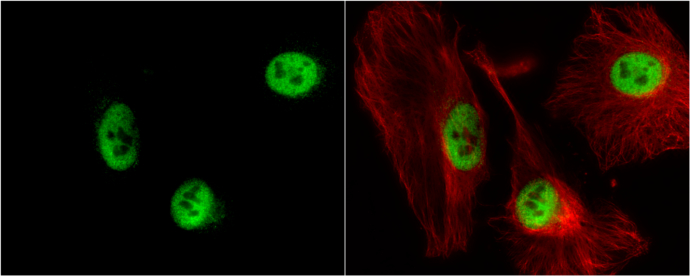
GTX115554 ICC/IF Image
RbAp48 antibody [N1C2] detects RbAp48 protein at nucleus by immunofluorescent analysis.
Sample: HeLa cells were fixed in 4% paraformaldehyde at RT for 15 min.
Green: RbAp48 protein stained by RbAp48 antibody [N1C2] (GTX115554) diluted at 1:500.
Red: alpha Tubulin, a cytoskeleton marker, stained by alpha Tubulin antibody [B-5-1-2] (GTX11304) diluted at 1:10000.
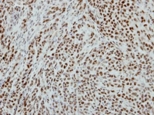
GTX115554 IHC-P Image
Immunohistochemical analysis of paraffin-embedded SCM1 xenograft, using RbAp48(GTX115554) antibody at 1:500 dilution.
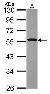
GTX115554 WB Image
Sample (30 ug of whole cell lysate)
A: JurKat
10% SDS PAGE
GTX115554 diluted at 1:3000
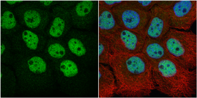
GTX115554 ICC/IF Image
RbAp48 antibody [N1C2] detects RbAp48 protein at nucleus by immunofluorescent analysis.
Sample: A431 cells were fixed in 4% paraformaldehyde at RT for 15 min.
Green: RbAp48 protein stained by RbAp48 antibody [N1C2] (GTX115554) diluted at 1:500.
Red: alpha Tubulin, a cytoskeleton marker, stained by alpha Tubulin antibody [GT114] (GTX628802) diluted at 1:1000.
Blue: Hoechst 33342 staining.
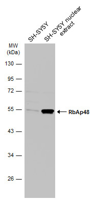
GTX115554 WB Image
SH-SY5Y whole cell and nuclear extracts (30 ug) were separated by 10% SDS-PAGE, and the membrane was blotted with RbAp48 antibody [N1C2] (GTX115554) diluted at 1:1000. The HRP-conjugated anti-rabbit IgG antibody (GTX213110-01) was used to detect the primary antibody.