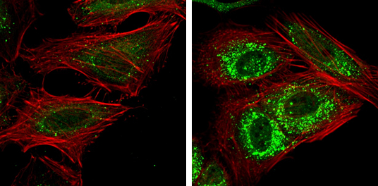
GTX115378 ICC/IF Image
NDP52 antibody detects NDP52 protein at autophagosome by immunofluorescent analysis.
Samples: HeLa cells mock (left) and treated with 50uM Chloroquine for 24 hr (right) were fixed in 4% paraformaldehyde at RT for 15 min.
Green: NDP52 protein stained by NDP52 antibody (GTX115378) diluted at 1:1000.
Red: Phalloidin, a F-actin marker.
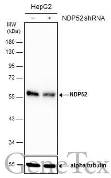
GTX115378 WB Image
Non-transfected (?) and transfected (+) HepG2 whole cell extracts (30 ug) were separated by 10% SDS-PAGE, and the membrane was blotted with NDP52 antibody (GTX115378) diluted at 1:4000. The HRP-conjugated anti-rabbit IgG antibody (GTX213110-01) was used to detect the primary antibody.
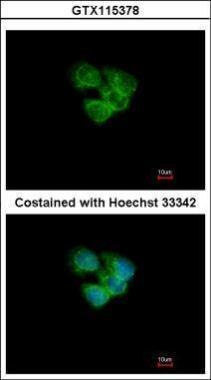
GTX115378 ICC/IF Image
Immunofluorescence analysis of methanol-fixed A431, using NDP52(GTX115378) antibody at 1:200 dilution.
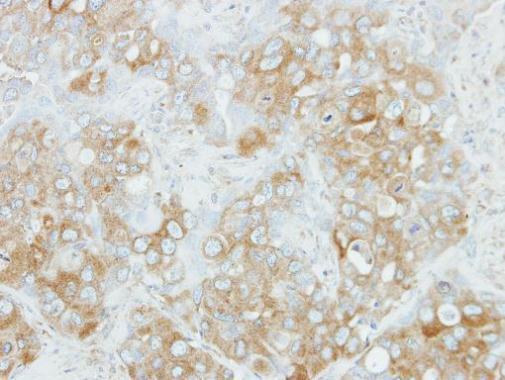
GTX115378 IHC-P Image
Immunohistochemical analysis of paraffin-embedded H441 xenograft, using NDP52(GTX115378) antibody at 1:100 dilution.
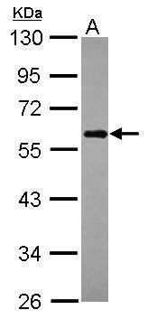
GTX115378 WB Image
Sample (30 ug of whole cell lysate)
A: Raji
10% SDS PAGE
GTX115378 diluted at 1:1000
The HRP-conjugated anti-rabbit IgG antibody (GTX213110-01) was used to detect the primary antibody.
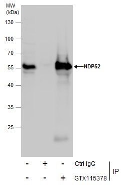
GTX115378 IP Image
Immunoprecipitation of NDP52 protein from Jurkat whole cell extracts using 5 ug of NDP52 antibody (GTX115378).
Western blot analysis was performed using NDP52 antibody (GTX115378).
EasyBlot anti-Rabbit IgG (GTX221666-01) was used as a secondary reagent.
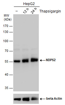
GTX115378 WB Image
NDP52 antibody detects NDP52 protein by western blot analysis. Un-treated (-) and treated (+, Thapsigargin treatment for 12hrs and 24hrs) HepG2 whole cell extracts (30 ug) were separated by 10% SDS-PAGE, and the membrane was blotted with NDP52 antibody (GTX115378) diluted by 1:2000.
The ACTB was used as internal control (GTX110564, 1:50000) shown at the bottom panel.
The HRP-conjugated anti-rabbit IgG antibody (GTX213110-01) was used to detect the primary antibody.
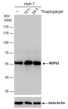
GTX115378 WB Image
NDP52 antibody detects NDP52 protein by western blot analysis. Un-treated (-) and treated (+, Thapsigargin treatment for 12hrs and 24hrs) Huh-7 whole cell extracts (30 ug) were separated by 10% SDS-PAGE, and the membrane was blotted with NDP52 antibody (GTX115378) diluted by 1:2000.
The ACTB was used as internal control (GTX110564, 1:50000) shown at the bottom panel.
The HRP-conjugated anti-rabbit IgG antibody (GTX213110-01) was used to detect the primary antibody.