
GTX115032 WB Image
Ataxin 3 antibody detects Ataxin 3 protein by western blot analysis.
293T mock transfected (-) or transfected with a construct expressing human Ataxin 3 (+).
10% SDS-PAGE
Ataxin 3 antibody (GTX115032) dilution: 1:5000
The HRP-conjugated anti-rabbit IgG antibody (GTX213110-01) was used to detect the primary antibody.
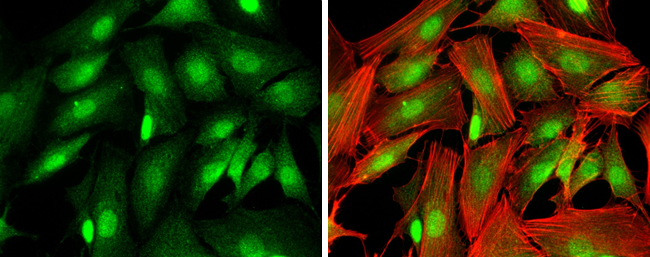
GTX115032 ICC/IF Image
Ataxin 3 antibody detects Ataxin 3 protein at cytoplasm and nucleus by immunofluorescent analysis.
Sample: SK-N-SH cells were fixed in 4% paraformaldehyde at RT for 15 min.
Green: Ataxin 3 protein stained by Ataxin 3 antibody (GTX115032) diluted at 1:250.
Blue: Hoechst 33342 staining.
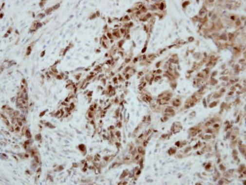
GTX115032 IHC-P Image
Immunohistochemical analysis of paraffin-embedded H661 xenograft, using Ataxin 3(GTX115032) antibody at 1:500 dilution.
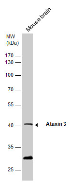
GTX115032 WB Image
Ataxin 3 antibody detects Ataxin 3 protein by western blot analysis. Mouse tissue extracts (50 ug) was separated by 10% SDS-PAGE, and blotted with Ataxin 3 antibody (GTX115032) diluted by 1:500. The HRP-conjugated anti-rabbit IgG antibody (GTX213110-01) was used to detect the primary antibody.
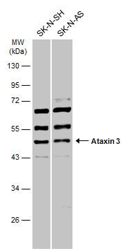
GTX115032 WB Image
Various whole cell extracts (30 ug) were separated by 10% SDS-PAGE, and the membrane was blotted with Ataxin 3 antibody (GTX115032) diluted at 1:1000. The HRP-conjugated anti-rabbit IgG antibody (GTX213110-01) was used to detect the primary antibody.

GTX115032 ICC/IF Image
Ataxin 3 antibody detects Ataxin 3 protein at cytoplasm and nucleus by immunofluorescent analysis.Sample: 293T cells were fixed in 4% paraformaldehyde at RT for 15 min.Green: Ataxin 3 stained by Ataxin 3 antibody (GTX115032) diluted at 1:500.Blue: Hoechst 33342 staining.