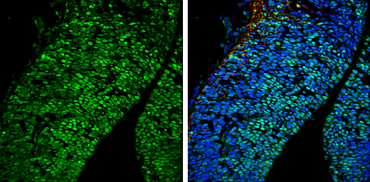
GTX114650 IHC-Fr Image
Brn2 antibody detects Brn2 protein expression at nucleus by immunohistochemical analysis.
Sample: Frozen sectioned E13.5 Rat brain.
Green: Brn2 protein stained by Brn2 antibody (GTX114650) diluted at 1:250.
Red: beta Tubulin 3/ TUJ1, a mature neuron marker, stained by beta Tubulin 3/ TUJ1 antibody [GT11710] (GTX631836) diluted at 1:500.
Blue: Fluoroshield with DAPI (GTX30920).
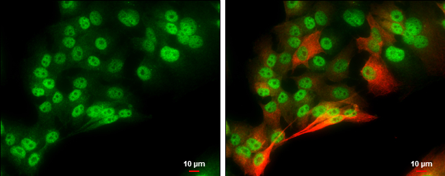
GTX114650 ICC/IF Image
Brn2 antibody detects Brn2 protein at nucleus by immunofluorescent analysis.
Sample: SK-N-AS cells were fixed in 4% paraformaldehyde at RT for 15 min.
Green: Brn2 protein stained by Brn2 antibody (GTX114650) diluted at 1:1000.
Red: beta Tubulin 3/ Tuj1, a cytoskeleton marker, stained by beta Tubulin 3/ Tuj1 antibody [GT11710] (GTX631836) diluted at 1:500.
Scale bar = 10 um.

GTX114650 ChIP assay Image
Brn2 antibody immunoprecipitates Brn2 protein-DNA in ChIP experiments. ChIP Sample: 293T whole cell lysate/extract A. 5 ug preimmune rabbit IgG B. 5 ug of Brn2 antibody (GTX114650) The precipitated DNA was detected by PCR with primer set targeting to MITF promoter.
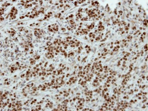
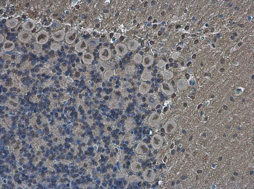
GTX114650 IHC-P Image
Immunohistochemical analysis of paraffin-embedded TOV-21G xenograft, using Brn2(GTX114650) antibody at 1:500 dilution.
GTX114650 IP Image
Brn2 antibody immunoprecipitates Brn2 protein in IP experiments. IP Sample: 1000 ug 293T whole cell lysate/extract A. 50 ug 293T whole cell lysate/extract B. Control with 2 ug of preimmune rabbit IgG C. Immunoprecipitation of Brn2 protein by 2 ug of Brn2 antibody (GTX114650) 10% SDS-PAGE The immunoprecipitated Brn2 protein was detected by Brn2 antibody (GTX114650) diluted at 1:1000. EasyBlot anti-rabbit IgG (GTX221666-01) was used as a secondary reagent.
GTX114650 IHC-P Image
Brn2 antibody detects Brn2 protein at nucleus in mouse brain by immunohistochemical analysis.
Sample: Paraffin-embedded mouse brain.
Brn2 antibody (GTX114650) diluted at 1:500.
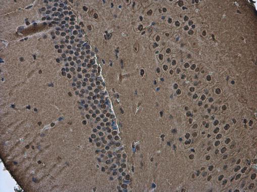
GTX114650 IHC-P Image
Brn2 antibody detects Brn2 protein at nucleus in rat brain by immunohistochemical analysis.
Sample: Paraffin-embedded rat brain.
Brn2 antibody (GTX114650) diluted at 1:500.
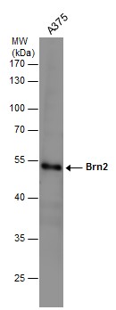
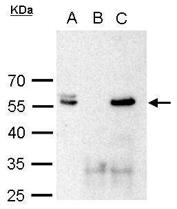
GTX114650 WB Image
Whole cell extract (30 ug) was separated by 10% SDS-PAGE, and the membrane was blotted with Brn2 antibody (GTX114650) diluted at 1:1000.