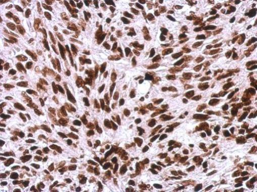
GTX114462 IHC-P Image
Immunohistochemical analysis of paraffin-embedded SkHep1 xenograft, using Histone H1.0(GTX114462) antibody at 1:500 dilution.
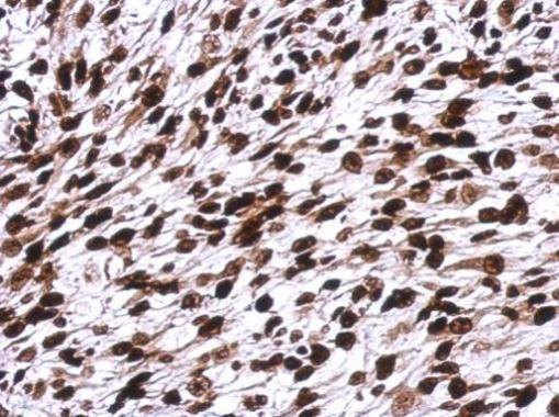
GTX114462 IHC-P Image
Immunohistochemical analysis of paraffin-embedded C2C12 xenograft, using Histone H1.0(GTX114462) antibody at 1:500 dilution.
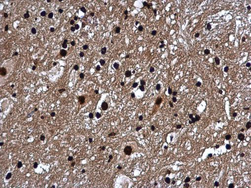
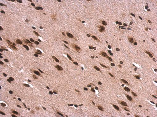
GTX114462 IHC-P Image
Histone H1.0 antibody detects Histone H1.0 protein at nucleus in mouse brain by immunohistochemical analysis.
Sample: Paraffin-embedded mouse brain.
Histone H1.0 antibody (GTX114462) diluted at 1:500.
GTX114462 IHC-P Image
Histone H1.0 antibody detects Histone H1.0 protein at nucleus in mouse brain by immunohistochemical analysis.
Sample: Paraffin-embedded mouse brain.
Histone H1.0 antibody (GTX114462) diluted at 1:500.
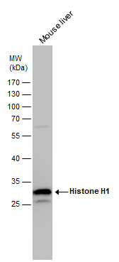
GTX114462 WB Image
Histone H1 antibody detects Histone H1 protein by western blot analysis. Mouse tissue extracts (50 ug) was separated by 12% SDS-PAGE, and the membrane was blotted with Histone H1 antibody (GTX114462) diluted by 1:5000. The HRP-conjugated anti-rabbit IgG antibody (GTX213110-01) was used to detect the primary antibody.
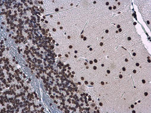
GTX114462 IHC-P Image
Histone H1.0 antibody detects Histone H1.0 protein at nucleus on rat fore brain by immunohistochemical analysis.
Sample: Paraffin-embedded rat fore brain.
Histone H1.0 antibody (GTX114462) dilution: 1:500.
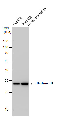
GTX114462 WB Image
Histone H1 antibody detects Histone H1 protein by western blot analysis. HepG2 whole cell extracts and nuclear extracts (30 ug) were separated by 12% SDS-PAGE, and the membrane was blotted with Histone H1 antibody (GTX114462) at a dilution of 1:10000 and developed with Trident femto Western HRP Substrate (GTX14698). The HRP-conjugated anti-rabbit IgG antibody (GTX213110-01) was used to detect the primary antibody.