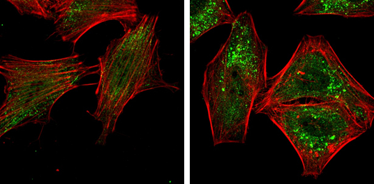
GTX113309 ICC/IF Image
ATG5 antibody detects ATG5 protein at autophagosome by immunofluorescent analysis.
Samples: HeLa cells mock (left) and treated with 50uM Chloroquine for 24 hr (right) were fixed in 4% paraformaldehyde at RT for 15 min.
Green: ATG5 protein stained by ATG5 antibody (GTX113309) diluted at 1:1000.
Red: Phalloidin, a F-actin marker.
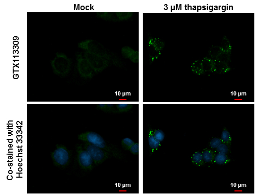
GTX113309 ICC/IF Image
ATG5 antibody detects ATG5 protein at autophagosome by immunofluorescent analysis.
Sample: HepG2 cells treated with 3uM thapsigargin 16 hrs (rigtht) and mock (left) and were fixed in ice-cold MeOH for 10 min, permeabilize with cooled acetone for 2 min for min.
Green: ATG5 protein stained by ATG5 antibody (GTX113309) diluted at 1:500.
Blue: Hoechst 33342 staining.
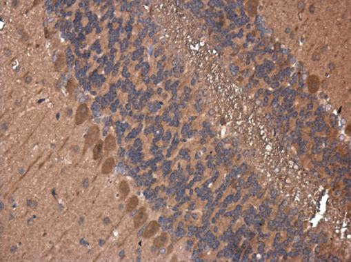
GTX113309 IHC-P Image
ATG5 antibody detects ATG5 protein at cytoplasm in mouse brain by immunohistochemical analysis.
Sample: Paraffin-embedded mouse brain.
ATG5 antibody (GTX113309) diluted at 1:400.
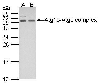
GTX113309 WB Image
Sample (30 ug of whole cell lysate)
A: NT2D1
B: PC-3
12% SDS PAGE
GTX113309 diluted at 1:1000
The HRP-conjugated anti-rabbit IgG antibody (GTX213110-01) was used to detect the primary antibody.
GTX113309 WB Image
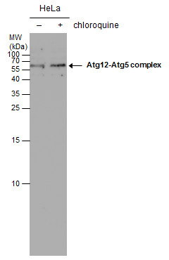
ATG5 antibody detects ATG5 protein by western blot analysis. Un-treated (-) and treated (+, 50 uM chloroquine treatment for 24 hrs) HeLa whole cell extracts (30 ug) were separated by 15% SDS-PAGE, and the membrane was blotted with ATG5 antibody (GTX113309) diluted at 1:1000. The HRP-conjugated anti-rabbit IgG antibody (GTX213110-01) was used to detect the primary antibody.