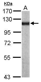
GTX113115 WB Image
Sample (50 ug of whole cell lysate)
A: Mouse brain
7.5% SDS PAGE
GTX113115 diluted at 1:1000
The HRP-conjugated anti-rabbit IgG antibody (GTX213110-01) was used to detect the primary antibody.
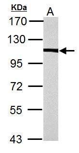
GTX113115 WB Image
alpha Actinin 4 antibody [N2C1], Internal detects alpha Actinin 4 protein by western blot analysis.
A. 30 ug PC-12 whole cell lysate/extract
7.5% SDS-PAGE
alpha Actinin 4 antibody [N2C1], Internal (GTX113115) dilution: 1:3000
The HRP-conjugated anti-rabbit IgG antibody (GTX213110-01) was used to detect the primary antibody.
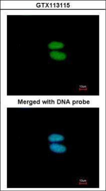
GTX113115 ICC/IF Image
Immunofluorescence analysis of paraformaldehyde-fixed HeLa, using alpha Actinin 4(GTX113115) antibody at 1:200 dilution.
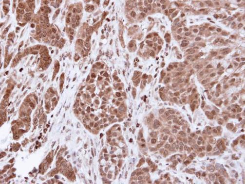
GTX113115 IHC-P Image
Immunohistochemical analysis of paraffin-embedded A549 xenograft, using alpha Actinin 4(GTX113115) antibody at 1:500 dilution.
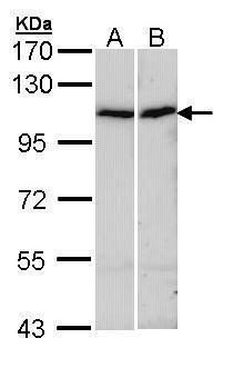
GTX113115 WB Image
Sample (30 ug of whole cell lysate)
A: A431 (GTX27909)
B: HepG2 (GTX27900)
7.5% SDS PAGE
GTX113115 diluted at 1:1000
The HRP-conjugated anti-rabbit IgG antibody (GTX213110-01) was used to detect the primary antibody.
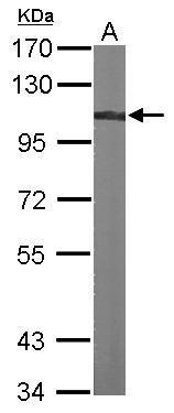
GTX113115 WB Image
Sample (30 ug of whole cell lysate)
A: NIH-3T3
7.5% SDS PAGE
GTX113115 diluted at 1:1000
The HRP-conjugated anti-rabbit IgG antibody (GTX213110-01) was used to detect the primary antibody.