GTX112981 IHC-Fr Image
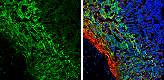
Tau antibody detects Tau protein expression by immunohistochemical analysis.
Sample: Frozen sectioned E13.5 Rat brain.
Green: Tau protein stained by Tau antibody (GTX112981) diluted at 1:250.
Red: beta Tubulin 3/ TUJ1, a mature neuron marker, stained by beta Tubulin 3/ TUJ1 antibody [GT11710] (GTX631836) diluted at 1:500.
Blue: Fluoroshield with DAPI (GTX30920).
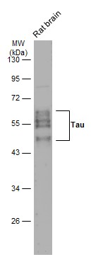
GTX112981 WB Image
Rat tissue extract (50 ug) was separated by 10% SDS-PAGE, and the membrane was blotted with Tau antibody (GTX112981) diluted at 1:1000. The HRP-conjugated anti-rabbit IgG antibody (GTX213110-01) was used to detect the primary antibody.
GTX112981 ICC/IF Image
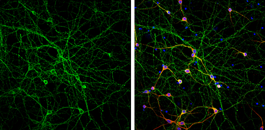
Tau antibody detects Tau protein at axon by immunofluorescent analysis.
Sample: DIV9 rat E18 primary cortical neurons were fixed in 4% paraformaldehyde at RT for 15 min.
Green: Tau protein stained by Tau antibody (GTX112981) diluted at 1:500.
Red: MAP2, stained by MAP2 antibody [HM-2] (GTX11267) diluted at 1:1000.
Blue: Fluoroshield with DAPI (GTX30920).
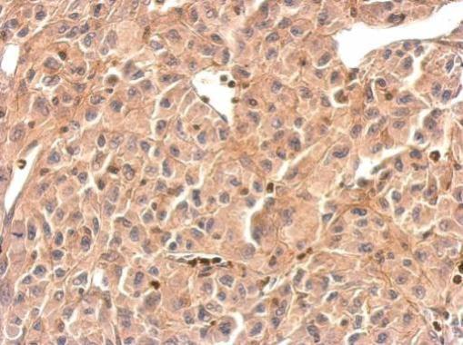
GTX112981 IHC-P Image
Tau antibody detects MAPT protein at cytosol on U87 xenograft by immunohistochemical analysis.
Sample: Paraffin-embedded U87 xenograft.
Tau antibody (GTX112981) dilution: 1:500.
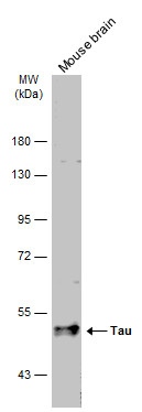
GTX112981 WB Image
Mouse tissue extract (50 ug) was separated by 7.5% SDS-PAGE, and the membrane was blotted with Tau antibody (GTX112981) diluted at 1:1000. The HRP-conjugated anti-rabbit IgG antibody (GTX213110-01) was used to detect the primary antibody.